Any extracts used in the following article are for non commercial research and educational purposes only and may be subject to copyright from their respective owners.
Background
Spike protein (inc vax) induced immunodeficiency & carcinogenesis megathread #21: BNT162b2 derived miR-21
https://doorlesscarp953.substack.com/p/spike-protein-inc-vax-induced-immunodeficiency-880
Quantum microRNA Assessment of COVID-19 RNA Vaccine: Hidden Potency of BNT162b2 SASR-CoV-2 Spike RNA as MicroRNA Vaccine
https://doorlesscarp953.substack.com/p/quantum-microrna-assessment-of-covid
SARS-CoV-2 Spike Targets USP33-IRF9 Axis via Exosomal miR-148a to Activate Human Microglia
https://doorlesscarp953.substack.com/p/sars-cov-2-spike-targets-usp33-irf9
What is fibrosis?
“The term fibrosis describes the development of fibrous connective tissue as a reparative response to injury or damage. Fibrosis may refer to the connective tissue deposition that occurs as part of normal healing or to the excess tissue deposition that occurs as a pathological process.”
https://www.news-medical.net/health/What-is-Fibrosis.aspx
The paper:
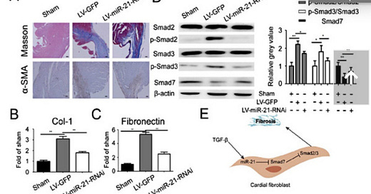
Mir-21 Promotes Cardiac Fibrosis After Myocardial Infarction Via Targeting Smad7 (2017)
Key Words: Mir-21 Cardiac fibrosis Myocardial infarction Smad7 TGF-β1
Abstract
Background/Aims: Cardiac fibrosis after myocardial infarction (MI) has been identified as an important factor in the deterioration of heart function. Previous studies have demonstrated that miR-21 plays an important role in various pathophysiological processes in the heart. However, the role of miR-21 in fibrosis regulation after MI remains unclear. Methods: To induce cardiac infarction, the left anterior descending coronary artery was permanently ligated of mice. First, we explored the expression of miR-21 in the infarcted zone in mice model of MI via RT-qPCR. Next, we examined the effects of TGF-β1 on miR-21 expression in cardiac fibroblasts (CFs). Then, CFs were infected with miR-21 mimics or miR-21 inhibitors to investigate the effects of miR-21 on the process of CFs activation in vitro. Further, bioinformatics analysis and luciferase reporter assay were performed to identify and validate the target gene of miR-21. At last, in-vivo study was done to confirm MiR-21 regulated myocardial fibrosis after MI in mice. Results: MiR-21 was up-regulated in the infarcted zone after MI in vivo. TGF-β1 treatment increased miR-21 expression in CFs. Overexpression of miR-21 promoted the effects of TGF-β1-induced activation of CFs, evidenced by increased expression of Col-1, α-SMA and F-actin, whereas inhibition of miR-21 attenuated the process of fibrosis. Bioinformatics, Western blot analysis and luciferase reporter assay demonstrated that Smad7 is a direct target of miR-21. In addition, in-vivo study revealed that MiR-21 regulated myocardial fibrosis after MI in mice. Conclusion: These findings suggested that miR-21 has a critical role in CF activation and cardiac fibrosis after MI through via TGF-β/Smad7 signaling pathway. Thus, miR-21 promises to be a potential therapy in treatment of cardiac fibrosis after MI.
Discussion
Cardiac fibrosis, which was an important part of cardiac remodeling, resulted in stiffening of the ventricular walls and diminished contractility and abnormalities in cardiac conductance, was a common consequence of numerous forms of heart diseases, including pathological hypertrophy, volume overload, and MI [37, 38].
In the past decade, numerous evidence has demonstrated the role of microRNAs in various pathophysiological processes, including cardiac fibrosis [17-20]. Among these fibrosis-related microRNAs, miR-21 was previously reported to play an important role in regulation of fibrosis in multiple tissues, such as kidney, liver and lung etc [25-27]. In the present study, we established mice models of cardiac fibrosis after MI and examined miR-21 expression at different time points and different cardiac zones.
Full paper:
https://www.karger.com/Article/FullText/479995
Pathology
I would regard it as contributory to the post BNT162b2 transfection sequalae.
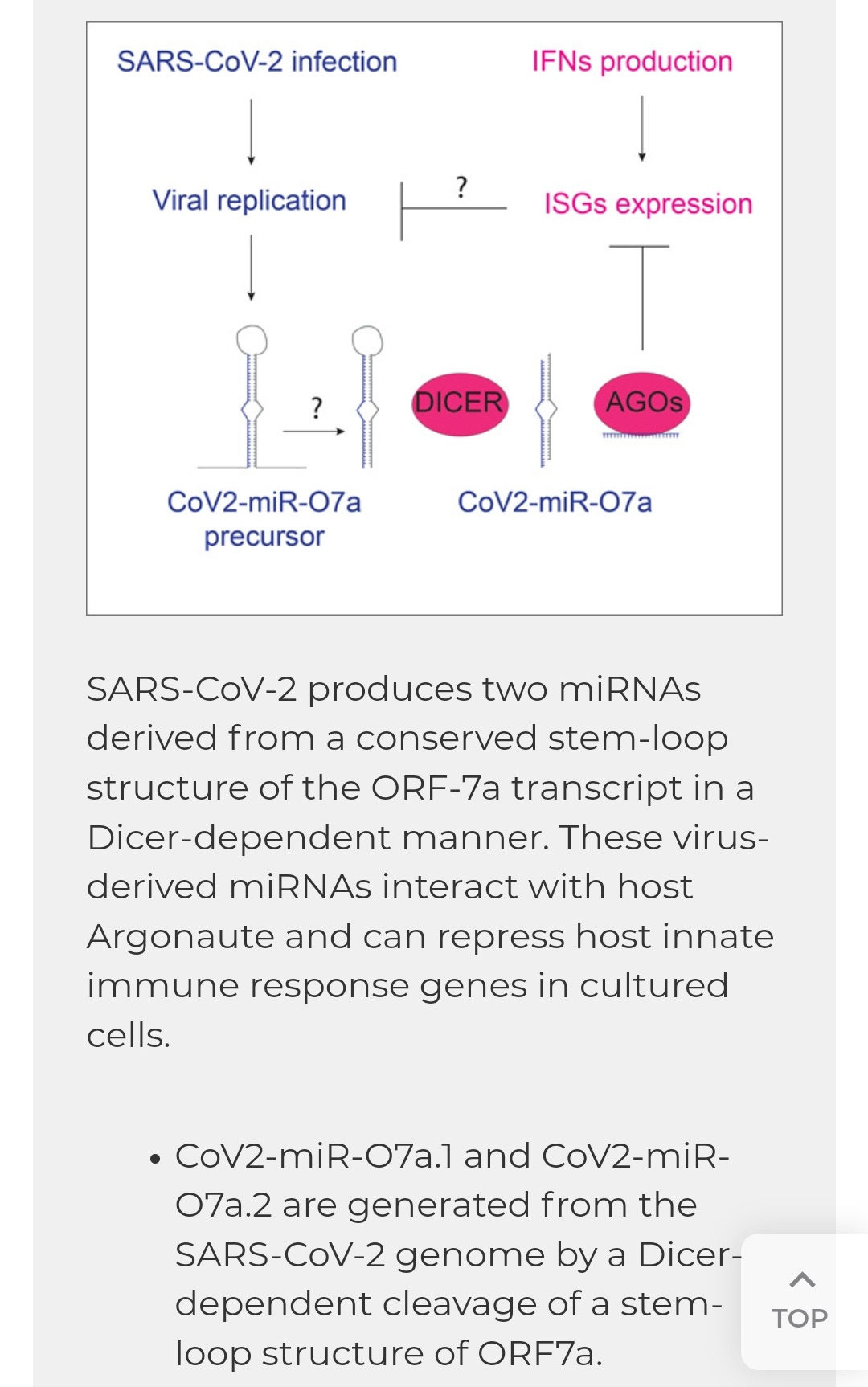
It appears to be specific to this agent, the virus itself also expresses microRNA's but these don't originate from the spike sequence and serve to repress host immunity.
Are we saying the therapeutic agent adds in a toxicity that even the virus itself lacked? MiR-21 is upregulated in response to severe infection but as far as we know it's not being expressed directly from the virus itself. Indeed it would appear so: gain of function.
Many other factors also contribute to cardiomyopathy, including ACE2 binding, cellular fusion, T-lymphocyte attack, ROS induced damage and so on. Fibrosis strongly hints at the risk of long term cumulative dose dependent damage, probably asymptomatic at first in most cases.
A virus-derived microRNA targets immune response genes during SARS-CoV-2 infection (2022)
Full paper:
https://www.embopress.org/doi/full/10.15252/embr.202154341
Myocardial Fibrosis as an Early Manifestation of Hypertrophic Cardiomyopathy (2010)
Abstract
BACKGROUND
Myocardial fibrosis is a hallmark of hypertrophic cardiomyopathy and a proposed substrate for arrhythmias and heart failure. In animal models, profibrotic genetic pathways are activated early, before hypertrophic remodeling. Data showing early profibrotic responses to sarcomere-gene mutations in patients with hypertrophic cardiomyopathy are lacking.
METHODS
We used echocardiography, cardiac magnetic resonance imaging (MRI), and serum biomarkers of collagen metabolism, hemodynamic stress, and myocardial injury to evaluate subjects with hypertrophic cardiomyopathy and a confirmed genotype.
RESULTS
The study involved 38 subjects with pathogenic sarcomere mutations and overt hypertrophic cardiomyopathy, 39 subjects with mutations but no left ventricular hypertrophy, and 30 controls who did not have mutations. Levels of serum C-terminal propeptide of type I procollagen (PICP) were significantly higher in mutation carriers without left ventricular hypertrophy and in subjects with overt hypertrophic cardiomyopathy than in controls (31% and 69% higher, respectively; P<0.001). The ratio of PICP to C-terminal telopeptide of type I collagen was increased only in subjects with overt hypertrophic cardiomyopathy, suggesting that collagen synthesis exceeds degradation. Cardiac MRI studies showed late gadolinium enhancement, indicating myocardial fibrosis, in 71% of subjects with overt hypertrophic cardiomyopathy but in none of the mutation carriers without left ventricular hypertrophy.
CONCLUSIONS
Elevated levels of serum PICP indicated increased myocardial collagen synthesis in sarcomere-mutation carriers without overt disease. This profibrotic state preceded the development of left ventricular hypertrophy or fibrosis visible on MRI. (Funded by the National Institutes of Health and others.)
Full paper:
https://www.nejm.org/doi/full/10.1056/nejmoa1002659
Pathogenesis and Prevention of Radiation-induced Myocardial Fibrosis (2017)
Abstract
Radiation therapy is one of the most important methods for the treatment of malignant tumors. However, in radiotherapy for thoracic tumors such as breast cancer, lung cancer, esophageal cancer, and mediastinal lymphoma, the heart, located in the mediastinum, is inevitably affected by the irradiation, leading to pericardial disease, myocardial fibrosis, coronary artery disease, valvular lesions, and cardiac conduction system injury, which are considered radiation-induced heart diseases. Delayed cardiac injury especially myocardial fibrosis is more prominent, and its incidence is as high as 20–80%. Myocardial fibrosis is the final stage of radiation-induced heart diseases, and it increases the stiffness of the myocardium and decreases myocardial systolic and diastolic function, resulting in myocardial electrical physiological disorder, arrhythmia, incomplete heart function, or even sudden death. This article reviews the pathogenesis and prevention of radiation-induced myocardial fibrosis for providing references for the prevention and treatment of radiation-induced myocardial fibrosis.
Keywords: RIHD, pathogenesis, cytokine, ROS, calcium overload, prevention
"Although RIHD subclinical abnormalities are progressive and found early, RIHD’s clinical symptoms appear 10 years after the end of radiotherapy."
Full paper:
https://www.ncbi.nlm.nih.gov/labs/pmc/articles/PMC5464468/
Case reports
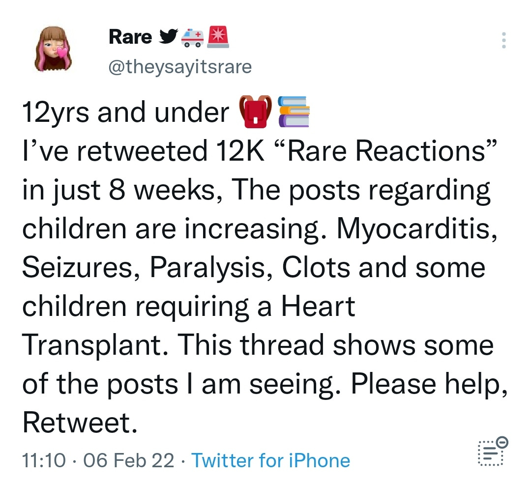
https://twitter.com/theysayitsrare/status/1490281616178036740?s=19
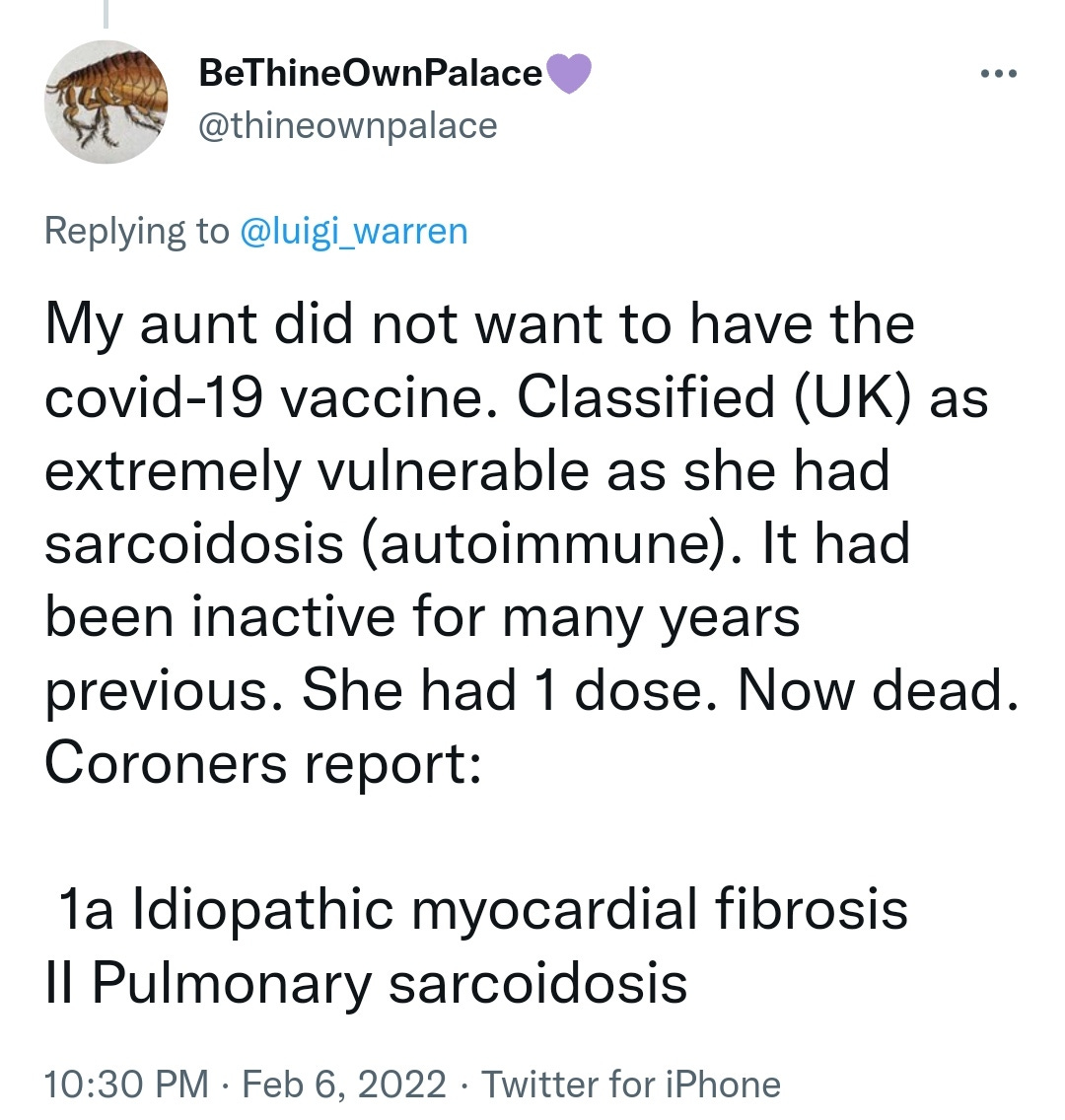
https://twitter.com/thineownpalace/status/1490452858172588037?s=20


