This Substack is too long for email. Please click on the title to load in the app or a browser.
Reading time:
short story - novelette - novella - novel - PhD thesis - War and Peace - U.S. Tax Code
Any extracts used in the following article are for non-commercial research and educational purposes only and may be subject to copyright from their respective owners.
It also discussed a secondary mechanism, whereby endogenous retroviruses could be inhibited or activated at the wrong time, and how this is linked to different cancers and diseases such as glioblastoma, heart disease, lupus, and arthritis.
This Substack will review some of the literature linking non-structural proteins to immunosuppression and progressive neurological diseases, and give an update on Kostaive side effects and its pending approval.
Discussion
Persistent expression and effects
Even if you don’t use other IIPs or nucleotide substitutions such as n1-methylpseudouridine (m1u) your innate immune signalling is still being suppressed by these products for as long as they are active. In a study using rabbits, saRNA from ATX-126 was still detectable in the liver, spleen, muscle and ovary at day 57.
This is long enough to initiate organ damage in the heart, kidneys, liver, pancreas, neurological disorders, autoimmune conditions, haematological disorders, and potential cancer progression or recurrence:
It’s a “feature” of replicon vaccines that I discussed in an earlier Substack:
“Severity also increased with dose and four (17%) of 24 participants who received 10.0μg reported a grade 3 (severe) systemic reaction after the firstor second vaccine.”
Between Dec 13, 2022, and Feb 25, 2023, we enrolled and randomly assigned 828 participants to receive ARCT-154 (n=420) or BNT162b2 (n=408) vaccines as a fourth-dose booster.
In per-protocol set 1, the GMTs of surrogate neutralising antibodies induced against the Wuhan-Hu-1 SARS-CoV-2 strain in the ARCT-154 group (5641 [95% CI 4321–7363]) were non-inferior to those in the BNT162b2 group (3934 [2993–5169]) when measured at 28 days after boosting, with a GMT ratio of 1·43 (95% CI 1·26–1·63).
Seroresponse rates were 65·2% (95% CI 60·2–69·9) in the ARCT-154 group versus 51·6% (46·4–56·8) in the BNT162b2 group, a difference of 13·6% (95% CI 6·8–20·5). GMTs against the omicron BA.4/5 variant on day 29 were 2551 (1687–3859) in the ARCT-154 group and 1958 (1281–2993) in the BNT162b2 group—a GMT ratio of 1·30 (1·07–1·58)—with seroresponse rates of 69·9% (65·0–74·4) and 58·0% (52·8–63·1).
Figure 3 Rates of solicited local reactions and systemic adverse events and their severity in the ARCT-154 and BNT162b2 groups
From: “Immunogenicity and safety of a booster dose of a self-amplifying RNA COVID-19 vaccine (ARCT-154) versus BNT162b2 mRNA COVID-19 vaccine: a double-blind, multicentre, randomised, controlled, phase 3, non-inferiority trial“ (2024)
And if you think that’s bad, an alarming study of transfected patients at 11 clinical sites in Japan found that neutralising antibodies persisted for at least 12 months after the last dose.
This is our second Lancet-published study. The irony is that the editors think they are supporting vaccines by sharing this efficacy data.
Serum antibodies are a poor correlate of immunity. They should be the “clean-up squad”, only called in where mucosal IgA, innate and adaptive responses have failed, and are virtually undetectable after 2-3 months.
The authors postulate that this was a “natural boosting” due to circulating infections, but I suspect that there is some persistent expression here, and we know that “long vaccine” syndrome has also been associated with this.
Either way, the effects are certainly “persistent”.
You can’t change your mind and there is no known antidote.
At 11 clinical sites in Japan, adults previously immunised with a minimum of three mRNA vaccine doses (last dose at least 3 months beforehand), were enrolled and randomly assigned to receive a booster dose of ARCT-154 or BNT16b2.3,4
Serum samples were obtained from all eligible participants at baseline before vaccination, and at 1, 3, 6, and 12 months post-vaccination to measure immunogenicity as neutralising antibodies against Wuhan-Hu-1 and Omicron BA.4-5 sub-lineage pseudoviruses.
… As previously reported,3,4 by 1 month both vaccines elicited increases in neutralising antibodies against both strains and to similar extents in both age groups (figure).
There were higher responses against Wuhan and Omicron BA.4-5 following ARCT-154 than BNT162b2, illustrated by GMT ratios of 1·45 (95% CI 1·22–1·71, Wuhan) and 1·31 (95% CI 1·01–1·71, Omicron BA.4-5) in the younger than 50 years group, and 1·42 (95% CI 1·18–1·72, Wuhan) and 1·29 (95% CI 0·96–1·73, Omicron BA.4-5) in the 50 years and older group.
These levels were essentially maintained in the ARCT-154 group to 3 months, while they had already started to decline in the BNT162b2 group.
Although GMTs eventually declined in both groups, the difference between groups increased over the 12 months of follow-up, with respective GMT ratios of 1·79 (95% CI 1·41–2·29) and 1·68 (1·15–2·45) for Wuhan and Omicron BA.4-5 in the younger than 50 years group, and 2·06 (1·55–2·75) and 2·14 (1·40–3·27) in the 50 years and older group (figure).
This superiority of ARCT-154 over BNT162b2 was supported by a consistent positive difference in seroresponse rates strains in both age groups (appendix p 3).
Figure Geometric mean neutralising antibody titres (with 95% CI) up to 12 months after vaccination with a booster dose of ARCT-154 or BNT162b2. Values are shown for titres against Wuhan-Hu-1 strain (A and B) and Omicron BA.4-5 variant (C and D) for participants grouped according to being age younger than 50 years (A and C) or 50 years and older (B and D). At each timepoint, numbers are n values for each group and the GMT ratio of titres between the two groups are given (with 95% CI). GMT=geometric mean titre.
… We also observed an increase in GMT against Omicron BA.2.86 in both vaccine groups from month 6 to month 12, suggesting some natural boosting of the response due to exposure to circulating virus (might be due to the dominant BA.2.86 and JN.1 lineages).8
We confirmed a better initial immune response against a panel of SARS-CoV-2 strains in preimmunised Japanese adults boosted with ARCT-154 compared with the conventional mRNA vaccine, BNT162b2, persisted up to 12 months post-vaccination, including in those age 50 years and older.
The observed superior immune response to ARCT-154 compared with BNT162b2 (measured by magnitude, persistence, and breadth) supports the preferential use of vaccines developed using self-amplifying mRNA technology over conventional mRNA vaccines as annual booster doses, but with updated formulations9 targeting recently emerged SARS-CoV-2 strains such as the JN.1 lineage to maintain protective immunity against emerging SARS-CoV-2 viruses.
From: “12-month persistence of immune responses to self-amplifying mRNA COVID-19 vaccines: ARCT-154 versus BNT162b2 vaccine“ (December 2024)
A preprint just published in medRxiv raises the bar even higher.
“Immunological and Antigenic Signatures Associated with Chronic Illnesses after COVID-19 Vaccination”, (18th February 2025)by Bhattacharjee et al1. reported finding detectable levels of circulating spike 709 days after COVID-19 mRNA injection. And it wasn’t rogue data, an isolated case.
PVS: post-vaccination syndrome.
Also known colloquially as Long Vaccine, or misdiagnosed as Long COVID or Postacute Sequelae of COVID-19 (PASC):
Blockbuster Yale Study: Millions Of Long COVID Patients Might Actually Be Vaccine Injured
Yale researchers released a study today that posits millions of Americans thought to have Long COVID may have been misdiagnosed and actually have post-vaccination syndrome caused by exposure to the spike protein in COVID vaccines.
The results indicated that participants with PVS had significantly higher circulating S1 levels compared with the control group (p = 0.01). However, circulating S1 was found in only a subset of participants with PVS at varying concentrations while the control group mostly exhibited a bimodal distribution of zero and non-zero values (Fig. 5A, Table S2).
Detectable S1 was found in participants’ plasma ranging from 26 to 709 days from the most recent known exposure (Figure 5B).
Non-structural proteins (nsPs) may also act as innate inhibiting proteins (IIPs)
Returning to the first Substack in the series, part 2 of the Japanese registration document specifies which nsPs are encoded by the saRNA used in the commercially available replicon vaccine ARCT-154 (“Kostaive”). (Emphasis mine throughout).
Note: They used Spike derived from the original, long-extinct strain of SARS-CoV-2. Even if it worked, this is a great way of priming your immune response to fail and to get breakthrough infections.
2. Quality and Outline of the Review Conducted by PMDA
2.1 Active substance
mRNA-2105 (active ingredient, zapomeran), an active substance of Kostaive, is a samRNA encoding replicase proteins (non-structural protein [nsP]1, nsP2, nsP3, and nsP4) of Venezuelan equine encephalitis virus (VEEV) origin and the full-length of S-protein (S1 and S2) derived from SARS-CoV-2 (of the original strain). mRNA-2105 also contains a 5’-terminal cap structure, 5’ untranslated region (UTR), UTR between reading frames, 3’UTR, and 3’-terminal poly A chain.
A paper from only four years reveals significant knowledge gaps, but helps to describe the mechanisms.
Who needs to fully understand how something works before mass-vaccinating the world?
Key takes from ”Alphavirus-Induced Membrane Rearrangements during Replication, Assembly, and Budding” (2021) by Elmasri et al.2
After uncoating, the genomic RNA (gRNA) is translated to yield two polyproteins. Of these polyproteins, ~10% contain nsPs 1, 2, 3, and 4 (P1234), while 90% contain nsPs 1, 2, and 3 (P123) [47].
After translation, the polyproteins containing nsP4 are processed by nsP2 to yield P123, as full-length P1234 is not capable of RNA synthesis [48].
P123 and nsP4 assemble into an early replication complex that facilitates genome replication. After its assembly, the early replication complex synthesizes negative-strand genomes using the positive-strand genome as a template [49].
Note that dsRNA intermediates are a necessary step:
Eventually, using the negative-strand and double-stranded RNA (dsRNA) intermediates, the replication complex synthesizes new copies of the +RNA genome [49].
We don’t know exactly how it works, but what the Hell, let’s use it commercially, and at scale:
Although structural information regarding most of the nsPs is available, because there is no known structure of the replication complex, the trigger that causes the switch from negative-strand to positive-strand synthesis is poorly understood.
However, it is believed that the eventual processing of P123 by nsP2 into individual nsP1, nsP2, and nsP3 irreversibly locks the replication complex into a late replication complex formation that is only capable ofsynthesizing +RNAs, specifically the gRNA and the subgenomic RNA (sgRNA) [47].
Note the role of dsRNA in the process. Of course, in a saRNA the viral organelles are not being generated, just the RNA copies and target protein(s) (e.g. full length Spike).
+RNA viruses hijack and rearrange cellular membranes into unique structures that support the replication of their genome [90,91]. These structures play a role in concentrating replicase components by spatially confining the replication process into specific compartments.
In addition, these structures shield replication intermediates such as dsRNA from host defense mechanisms [92].
From: “Figure 1.Alphavirus-induced replication organelles. Alphavirus replication spherules form at the plasma membrane and require the presence of nsP1, nsP2, nsP3, and nsP4. Structural studies proposed that the nsP1 ring complex forms the base of the alphavirus RC and plays a role in membrane curvature. The HVD of nsP3 contains a YXXM motif capable of binding to p85, relieving p110 inhibition. P110 catalyzes the phosphorylation of PIP2, generating PIP3, which can recruit and activate Akt at the plasma membrane. Active Akt promotes spherule internalization from the plasma membrane in an actin-myosin-dependent manner. Endocytic spherule-containing vesicles fuse with late endosomes to form acidic vesicles that traffic to the perinuclear area via microtubules, where they mature to form large CPV-Is. Figure created with BioRender.com.” Source: https://www.mdpi.com/2076-0817/10/8/984
Even without the viral organelles, a significant amount of the viral genome is being used in ARCT-154:
From: “Figure 1.Proteins identified to associate with CHIKV nsP3 HVD. (A) The organization of CHIKV genome, the domains of nsP3 and schematics of constructs for expressing EGFP-CHIKVnsP3HVD and non-tagged EGFP in Flp-In T-REx cells; (B) Immunoblot analysis using anti-EGFP antibodies and immunoprecipitated (IP) samples obtained from the parental Flp-In T-Rex cells and transgenic T-REx-EGFP and T-REx-EGFP-CHIKVnsP3HVD cell lines; (C) Interaction network of proteins associated with the HVD of CHIKV nsP3 was created using STRING database; for this analysis proteins that showed at least 2-fold enrichment in SILAC-based quantitative assay were chosen.” Source: https://www.mdpi.com/1999-4915/10/5/226
In 2022, Liu et al. published a very useful reference, the review article “Innate immune evasion by alphaviruses”3.
The paper covers all the key areas that alphaviruses use to escape immunosurveillance. These include “Restraint of cGAS-STING pathway”, “Inhibition of IFN pathway”, “Suppression of transcriptional host shutoff pathway”, “Suppression of translational host shutoff pathway”, and “Inhibition of RNAi pathway”.
Focussing on nsPs, this is what they have to say about the dual role of these proteins:
"... nsP2 from old world alphaviruses, including SINV and SFV, is the key regulator of the interaction between the virus and the host cell.
Not only does nsP2 serves as a component of the replicase complex required for viral RNA replication and transcription, but also it can directly participate in the inhibition of host transcription (48, 49). "
Viruses let very little go to waste.
To the vaccinee, almost every alphavirus protein is working to suppress their immune system:
Almost all alphavirus proteins antagonize innate immunity in different mechanisms and degrees.
The role of nsP1
BST-2: Bone marrow stromal antigen 2.
NsP1 downregulates the expression of BST-2, which acts as a well-characterised antiviral interferon-stimulated gene (ISG).
Recall that ARCT-154 encodes for nsPs 1-4 inclusive, from Venezuelan equine encephalitis virus (VEEV).
Through the interaction with cGAS-STING, nsP1 degrades cGAS to stabilize the virus protein (Figure 1). nsP1 also down-regulates the expression of BST-2 to inhibit the adhesion of VLPs on the plasma membrane and promote the release of the virus.
There is a lot happening here, but the part to focus on first is at the top-middle of the figure.
The red T-lines from E1, nsP2, and E2 indicate blocking of the PRRs RIG-I and MDA5 and downstream IFN-β signalling pathways.
This also blocks tumour-associated immune signalling.
Middle-left shows nsP4, nsP2 (and capsid from a live virus) causing cGAS degradation.
NsP1 is blocking STING, and on the right both nsP1 and nsP2 are blocking interferon-stimulated gene pathways:
NsP2 inhibits IFN type 1 signalling via the JAK-STAT pathway.
The mechanism of nsP2 antagonizing immune response is more complex. The type I interferon response can be counteracted by inhibiting the general transcription of host cells and reducing the phosphorylation of STAT1 in the JAK-STAT pathway (Figure 1).
RPB1 (= POLR2A) is a polymerase. It’s an essential component for transcribing DNA into mRNA.
Ubiquitination is the process whereby nsP2 targets it for destruction:
In addition, degradation of RPB1 occurs by nsP2-mediated ubiquitination, through which the transcription of the host proteins can be shut down (Figure 2). At the same time, nsP2 exerts a profound impact on the phosphorylation of STAT1 and STAT2 and thus inhibits the host translation (Figure 1).
NsPs 3 and 4 may also induce host transcriptional shutdown. NsP4 inhibits phosphorylation of eukaryotic translational initiation factor 2 (eIF2alpha).
The IIP K3 also targets this pathway (discussed in the last Substack).
Both nsP3 and nsP4 could induce the host transcriptional shutdown (Figure 2). NsP4 inhibits the phosphorylation of eIF2alpha in the PERK pathway.
Among the structural proteins, the capsid protein can effectively inhibit RNAi in insect and mammalian cells by separating double-stranded RNA and small interfering RNA (Figure 3).
E1 and E2 act as IIPs via inhibition of the pattern recognition receptors MDA5/RIG-I andinterferon beta signalling.
TF is a viral transferase protein.
E2 and E1 inhibit the activation of the IFN-β promoter induced by the MDA5/RIG-I receptor signaling pathway. TF antagonizes the host interferon response.
ZAP: a zinc-finger antiviral protein, which is stimulated by IFN. It inhibits the replication of some viruses, but some alphaviruses have evolved to evade ZAP recognition.
ZAP has been found to target three 500-bp sequences in alphavirus nsP2 RNA4. Have these sequences also been retained in ARCT-154? This would be unfortunate, as studies have shown that it also acts as a tumour suppressor in colorectal cancer (CRC)5. For the people of Japan, I wish that I was making this stuff up …
In addition to the structural and nonstructural proteins of alphavirus, the virus also uses the immune escape phenomenon of ZAP to antagonize the host’s antiviral response.
As more and more studies are performed, a deeper and more comprehensive understanding of alphavirus antagonizing host antiviral innate immunity is revealed. However, some mechanisms are not clear enough, and there may be other ways and mechanisms to antagonize antiviral immunity that are worthy of further research and exploration.
Here we report ZAP as a genuine tumor suppressor. Pan-can[c]er analysis with TCGA data from 712 patients and large-scale immunohistochemistry in tissue microarrays from 1552 patients reveal that ZAP is prevalently downregulated, and associated with poor survival in liver, colon, and bladder cancer patients.
Ectopic over-expression of ZAP inhibits the malignant phenotypes of colorectal tumor by cell cycle arrest. Using RNA immunoprecipitation and RNA decay assays, we demonstrate that ZAP directly and specifically binds to and degrades the transcript of TRAILR4, which in turn represses TRAILR4 expression and inhibits the aggressiveness of colorectal cancer cells.
Furthermore, our CRISPR-engineered mice models show that loss-of-function of ZAP synergizes with APC-deficiency to drive malignant colorectal cancer in vivo. Overall, we identify a previously unknown function of the antiviral factor ZAP in colorectal tumorigenesis, linking intrinsic immunity to tumor pathogenetics.
From: “Zinc-finger antiviral protein acts as a tumor suppressor in colorectal cancer“ (2020)
ARCT-154 replicase protein, encephalitis and multiple sclerosis (MS)
As if the science behind Kostaive couldn’t get any darker, Dr Stebel sent me a partly redacted snippet from the registration documents.
Why did they need to use amino acid substitutions in the replicase protein (nsPs)? What “cytotoxic effects” do they mean, and why is it redacted?
Note the weasel words: “reduces the cytotoxic effect of the replicase protein”, i.e.not eliminated.
S-protein has undergone substitution of 6 amino acid residues (D614G, R682G, R683S, R685S, K986P, and V987P) for enhanced immunogenicity. The replicase protein has undergone * amino acid substitutions (****** at **** and ****** at ****). Mutation of ****** at **** reduces the cytotoxic effect of the replicase protein, thereby contributing to the prolongation of S-protein expression. The mutation of ****** at **** allows constitutive expression of ****, enhancing RNA-replicating efficacy and contributing to an increase in S-protein expression level.
Dr. Stebel: “In Kostaive they also encode a modified replicase Enzyme. Is there any data of this protein in comparison to the wild type?”
To try to get some answers, this meant another literature review, as the regulators don’t appear to want us to know.
TL;DR:I followed several papers cited from 2000 to 1990. Neuropathology and death of mice were associated with wild type (wt)nsP3, a replicase component. Are they concerned about inducing autoimmune CNS damage?
Quick takes from five papers in chronological order from 1990 - 2021.
ELI5: Alphavirus infection of cells which protect nerves with a protective myelin sheath, called oligodendrocytes, leads to autoimmune attack, and this initiates progressive neurological diseases resembling MS.
From “Pathogenicity of Semliki Forest virus for the rat central nervous system and primary rat neural cell cultures: possible implications for the pathogenesis of multiple sclerosis” (1990) by Atkins et al6. (Paywalled beyond abstract).
The neurovirulent L10 strain of Semliki Forest virus (SFV) causes extensive neuronal damage in the central nervous system (CNS) of infected rats, and is probably the cause of death.
“Avirulent” means that the strains are not virulent or pathogenic - not capable of causing disease - but these findings contradict this:
The avirulent A7 and M9 strains do not cause extensive neuronal damage, but do induce immune-mediated CNS demyelination. In primary CNS cell cultures derived from rats, L10 multiplies more rapidly in neurons than avirulent strains, but infection with both virulent and avirulent strains causes depletion of oligodendrocytes from mixed glial cell cultures.
It is proposed that the immune-mediated demyelination, which follows infection with avirulent strains, is induced by phagocytosis of myelin debris from infected oligodendrocytes, and the presentation of antigens derived from such debris to T-helper lymphocytes.
Based on these and previous results, a scheme for the pathogenicity of defined strains of SFV is proposed. The applicability of this scheme to the understanding of human demyelinating disease such as multiple sclerosis is discussed.
Ten years on, and in this paper the authors discuss the effects of infecting rodents with different alphavirus nsP strains:
From “Replicase Complex Genes of Semliki Forest Virus Confer Lethal Neurovirulence”(2000) by Tuittila et al.7:
Semliki Forest virus (SFV) is a mosquito-transmitted pathogen of small rodents, and infection of adult mice with SFV4, a neurovirulent strain of SFV, leads to lethal encephalitis in a few days, whereas mice infected with the avirulent A7(74) strain remain asymptomatic.
In adult neurons, A7(74) is unable to form virions and hence does not reach a critical threshold of neuronal damage.
A smoking gun for nsP-mediated autoimmune disorders:
We first localized virulence determinants in the nonstructural region by showing that rA774 structural genes combined with the SFV4 nonstructural genome produced a highly virulent virus, while a reciprocal recombinant was asymptomatic.
In addition to several amino acid mutations in the nonstructural region, the nsp3 gene of rA774 displayed an opal termination codon and an in-frame 21-nucleotide deletion close to the nsp4 junction.
Replacement in rA774 of the entire nsp3 gene with that of SFV4 reconstituted the virulent phenotype, whereas an arginine at the opal position significantly increased virulence, leading to clinical symptoms in mice.
Completion of the nsp3 deletion in rA774 did not increase virulence.
Pathogenicity is reduced by using amino acid substitutions in the nsP:
We conclude that the opal codon and amino acid mutations other than the deleted residues are mainly responsible for the attenuation of A7(74) and that the attenuating determinants reside entirely in the nonstructural region.
The neurotoxic opal codon is also in VEEV, which ARCT-154 uses:
In several alphaviruses, such as SIN, Middelburg virus (43), and Ross River virus (42), as well as Venezuelan (16) and western and eastern (48) equine encephalitis viruses, an opal (UGA) termination codon interrupts the polygenic RNA at the 3′ end of the nsp3 gene.
Although in many alphaviruses mutations in the virion proteins or nucleocapsid have been found to alter virulence (11, 36), often having a synergistic effect (32), the results presented in this paper strongly suggest that the replicase complex nsp3 gene is the main pathogenic determinant conferring the avirulent phenotype of A7(74) and provide a rare example of the presence of an opal termination codon in one alphavirus strain but not in another.
Whether viral or not in origin, you don’t want a concentration of these nsPs around your CNS or brain:
It has been suggested that virulent strains reach a critical threshold titer in the central nervous system (CNS), leading to neuronal damage by either necrosis or apoptosis (10, 37).
You can understand why the regulators redacted the section and didn’t want to discuss the risks of neurotoxicity:
The chimeras obtained were designated rA774-V4nsp12 and rA774-V4nsp34, respectively (Fig. 1). rA774-V4nsp12 showed increased virulence, killing 7 of 20 mice (Fig. 3) and causing paralytic symptoms in an additional 6 animals, proving the involvement of this region in regulation of pathogenicity.
rA774-V4nsp34 replicated to high titers in the CNS of mice (Fig. 2) and killed 95% of the infected animals (Fig. 3). As sequence analysis had revealed major modifications in nsp3, the effect on virulence of this gene was analyzed next.
A highly neurovirulent virus with a lethal phenotype comparable to that of parental SFV4 and rA774-V4nstr was obtained by replacement of the nsp3 gene in rA774 with the corresponding SFV4 nsp3 gene.
The phenotype of this recombinant, designated rA774-V4nsp3 (Fig. 1), was confirmed by histological analysis, and the virus showed wide spreading in the cortex and cerebellum, with Purkinje neurons and granule cells strongly affected (Fig. 4C; also see below).
From “FIG. 4. SFV polyclonal antibody-stained brain sections from mice infected i.p. with 106 PFU of virus. (A) rA774 infection. Multiple small antigen-positive foci of virus are visible in the pons (P) and cerebellum (C) (arrows). Bar, 0.5 mm. (B) Widespread rA774-V4nstr infection of the pons (P), indicated with asterisks. Arrows, infected granule cells and cerebellar (C) Purkinje neurons with antigen-positive dendrites. Bar, 0.7 mm. (C) rA774-V4nsp3-infected areas of pons (P, asterisk) and cerebellum (C) with affected Purkinje cells, including dendrites (arrows). Bar, 0.6 mm. (D) rA774-V4del-infected cerebellum with a few viral foci (arrows). Bar, 0.6 mm. (E) Extensive rA774-arg invasion (asterisk) of the cerebral nucleus (putamen). Bar, 0.6 mm. (F) Widespread infection with V4-opal of brain hemisphere (arrows) of a mouse infected with the V4-opal construct which reverted to arginine. Bar, 0.7 mm.” Source: https://pmc.ncbi.nlm.nih.gov/articles/PMC111978/
They also investigated the effects of different opal variants on mice:
Both moribund mice sampled in this group were shown to be infected with opal revertants, and they displayed extensive virus spreading. The arginine revertant shown in Fig. 4F invaded the cortex and cerebrum.
This mouse was healthy, apart from having a paralysed left hind leg (!):
We also undertook histological studies on one affected mouse from the mortality study group, indicated with an asterisk in Fig. 3, which displayed left hind limb paralysis although otherwise clinically healthy.
Macrophage invasion of the spinal cord, associated with phenomena similar to Wallerian degeneration:
Severe myelin destruction was manifested, with numerous macrophagesin the spinal cord, both phenomena similar to those observed during Wallerian degeneration (not shown).
Wallerian degeneration is an active process of anterograde degeneration of the distal end of an axon that is a result of a nerve lesion. It occurs between 7 to 21 days after the lesion occurs. After the 21st day, acute nerve degeneration will show on the electromyography.
Presentations of nerve damage may include:
Reduced or loss of function in associated structures to damaged nerves
Gradual onset of numbness, prickling or tingling in feet or hands, which can spread upward into legs and arms
Sharp, jabbing, throbbing, freezing, or burning pain
The opal codon in nsp3 of rA774 was an unexpected finding, because the SFV4 and CA7 strains (46) both express an arginine codon at this site.
Although most alphaviruses express an opal codon (16, 42, 43, 48), its significance in the regulation of alphavirus pathogenesis remains elusive.
Such human pathogens as the Venezuelan (16) and eastern (48) encephalitis viruses, which cause severe disease, contain an opal codon at this position, indicating its selective advantage and stability.
The authors conclude that the opal site in nsp3 contributed to the pathogenicity:
The present study shows that in SFV, the opal site in nsp3 affects virus pathogenesis in mice. The introduction of an opal codon dramatically attenuated SFV4, making it nearly avirulent for adult mice, while replacement of the opal codon in rA774 with an arginine clearly increased pathogenicity even if it was not sufficient to restore full virulence.
While SFV4, rA774-V4nstr, and rA774-V4nsp3 all caused neurological symptoms within a short period before the mice succumbed, the rA774-arg mutant, which killed only one mouse, nevertheless frequently caused transient paralysis and limb weakness, thus exhibiting a medium degree of virulence not observed for rA774.
NsP1 and nsP2 are also implicated in contributing to neurotoxicity:
Although the contribution to pathogenicity of the nsp1 gene was not separately tested, it probably is involved, because in a reciprocal analysis, an SFV4 derivative expressing nsp1 of rA774 displayed a remarkably attenuated phenotype in adult mice (unpublished data). This does not, however, exclude the involvement of nsp2as well, because a few amino acid mutations were found in this region (Table 1).
The following year, Vihinen et al. sought to identify which amino acids on nsP3 were responsible for pathogenicity in mice, and how to reduce these effects. They found that it wasn’t so much the protein itself as phosphorylation by native cellular kinases (i.e. protein activation) that was responsible.
If you are developing a replicon vaccine how can you predict these effects (or not) in a genetically and phenotypically diverse population?
How can you do this in the new target of 100 days?
From “Elimination of Phosphorylation Sites of Semliki Forest Virus Replicase Protein nsP3*“ (2001)8:
nsP3 is one of the four RNA replicase subunits encoded by alphaviruses. The specific essential functions of nsP3 remain unknown, but it is known to be phosphorylated on serine and threonine residues.
Experiments with deletion variants suggested that nsP3 itself had no kinase activity; instead, it was likely to be phosphorylated by multiple cellular kinases.
Phosphorylation was not necessary for the peripheral membrane association of nsP3, which was mediated by the N-terminal region preceding the phosphorylation sites.
Mutations do have an association with rates of RNA synthesis, but the previous paper found exceptions to the rule:
Two deletion variants of nsP3 with either reduced or undetectable phosphorylation were studied in the context of virus infection. Cells infected with mutant viruses produced close to wild type levels of infectious virions; however, the rate of viral RNA synthesis was significantly reduced in the mutants.
If you want a high rate of RNA synthesis these tend to be associated with more pathogenic mutations:
A virus totally defective in nsP3 phosphorylation and exhibiting a decreased rate of RNA synthesis also exhibited greatly reduced pathogenicity in mice.
Our next paper from 2016 has a title that would explain the news blackout and censorship: “Alphavirus Encephalomyelitis: Mechanisms and Approaches to Prevention of Neuronal Damage” by Griffin9:
Epigenetic factors such as age, as well as genetic background, help to determine your risk of developing disorders such as MS after vaccination.
There is a reason why your risk increases with age. (N.B. most people only get shingles once. You might say its “self vaccinating”. If you are “severely immunocompromised” then a vax is a poor choice. Note the corporate logo and the pharma takeover of the NHS.) Source: https://www.getshinglesready.co.uk/resources/
Mosquito-borne viruses are important causes of death and long-term neurologic disability due to encephalomyelitis. Studies of mice infected with the alphavirus Sindbis virus have shown that outcome is dependent on the age and genetic background of the mouse and virulence of the infecting virus.
Age-dependent susceptibility reflects the acquisition by neurons of resistance to virus replication and virus-induced cell death with maturation.
In mature mice, the populations of neurons most susceptible to infection are in the hippocampus and anterior horn of the spinal cord. Hippocampal infection leads to long-term memory deficits in mice that survive, while motor neuron infection can lead to paralysis and death.
It’s not the virus itself, but the immune response that causes the damage (plus adverse events)
Neuronal death is immune-mediated, rather than a direct consequence of virus infection, and associated with entry and differentiation of pathogenic T helper 17 cells in the nervous system.
To modulate glutamate excitotoxicity, mice were treated with an N-methyl-D-aspartate receptor antagonist, α-amino-3-hydroxy-5-methyl-4-isoxazole propionic acid receptor antagonists or a glutamine antagonist.
Motor neurons (muscle firing) were at a greater risk than hippocampal neurons in the brain:
The N-methyl-D-aspartate receptor antagonist MK-801 protected hippocampal neurons but not motor neurons, and mice still became paralyzed and died. α-Amino-3-hydroxy-5-methyl-4-isoxazole propionic acid receptor antagonists GYKI-52466 and talampanel protected both hippocampal and motor neurons and prevented paralysis and death.
Your memory may also be affected if you react badly:
Glutamine antagonist 6-diazo-5-l-norleucine protected hippocampal neurons and improved memory generation in mice surviving infection with an avirulent virus.
You see the problem here. The whole point of using replicon vaccines such as ARCT-154 is to generate an immune response, and the use of nsPs (and dsRNA) is integral to their functionality.
The objective is to generate as much immunogenic protein as possible, for as long as possible.
Surprisingly, in all cases protection was associated with inhibition of the antiviral immune response, reduced entry of inflammatory cells into the central nervous system, and delayed virus clearance, emphasizing the importance of treatment approaches that include prevention of immunopathologic damage.
Delving more into the mechanisms: IFN pathways, T cell signalling and macrophage infiltration are implicated, as is the balance between pro-inflammatory cytokines and anti-inflammatory IL-10:
Several observations have led to the conclusion that neuronal damage in mature animals is primarily due to the antiviral immune response rather than virusreplication per se, and that fatal alphaviral encephalomyelitis is a T-cell-mediated immunopathologic process.
When you start to feel better, you start to feel worse:
For instance, initiation of virus clearance and the inflammatory response are coincident with the onset of neurological disease [21], and survival is improved in mice deficient in αβ T cells, β2-microglobulin, transporter associated with antigen processing (TAP), or CD4 but not in mice deficient in production of antibody, CD8, perforin, Fas, TNF-α receptor-1, IFN-γ, IFN-γ receptor-1, or IL-6 [34–36].
The virus has been cleared, yet mice still succumbed to Hydrocephalus ex vacuo, (also known as compensatory enlargement of the CSF spaces). This is usually seen with conditions that promote brain shrinkage in humans, such as Alzheimer’s disease, leukodystrophies, stroke or traumatic injuries10:
Furthermore, mice protected from fatal disease by passive transfer of immune serum after NSV infection clear infectious virus but develop a progressive loss of parenchyma (ex vacuo hydrocephalus) associated with infiltration of CD4+ T cells and macrophages into the hippocampus [35].
IL-10 is an important regulatory cytokine that helps to determine the balance between inflammation and immunoregulation [37, 38]. Deficiency of IL-10 accelerates the onset of fatal NSV-induced paralytic disease with an early increase in the CNS of CD4+ T cells expressing the transcription factor RORγt and producing the cytokine IL-17 [T helper (Th) 17 cells] [39].
Cytotoxic T lymphocytes kill virus-infected and tumour cells with a mediator of allergic inflammation called granzyme B:
Th17 cells are multifunctional and can have pathogenic or nonpathogenic characteristics. In response to NSV infection, Th17 cells in the CNS (but not in the draining lymph nodes) had a pathogenic phenotype with production of granulocyte macrophage colony-stimulating factor (GM-CSF) and granzyme B.
“Pathogenic” is good, “doubly pathogenic” is better. it must be the motivation behind replicon vaccines.
Note: T-bet helps to induce endogenous IFNgamma (IFN-γ) production. It also represses anti-inflammatory IL-4 levels and suppresses IL-511. IL-5 mediates eosinophilic inflammation and stimulates B cell growth:
In addition, some Th17 cells in the CNS developed intodoubly pathogenicTh1/Th17 cells withadditional expression of the transcription factor T-bet and production of IFN-γ.
Although pathogenic Th17 cells are recognized to be effectors in autoimmune disease [40], they were not previously identified as contributors to virus-induced immunopathology [41].
Low levels of IL-10 were associated with increased mortality, due to CD4+ T cell infiltration of the brain after alphavirus infection. These do not originate in the CNS:
These studies and comparative studies of BALB/c mice that are genetically resistant to fatal NSV-induced encephalomyelitis indicate the importance of IL-10 in regulating the immunopathogenic effects of antiviral T cells [42].
CD4+ T cells infiltrating the brains of BALB/c mice include fewer Th17 cells and more regulatory T cells producing IL-10 than similarly infected C57BL/6 mice [42].
In the absence of IL-10 BALB/c mice become susceptible to fatal infection.The primary sources of regulatory IL-10 during infection are the infiltrating CD4+ and CD8+ lymphocyte populations, not myeloid cells intrinsic to the CNS [43].
Naturally, the mechanisms are little understood. I guess there is no need to have mastery of your subject before authorising a mass-vaccination campaign:
Determination of the role of Th17 cells in NSV-induced immunopathology and identification of the mechanism(s) by which they influence outcome will be important for developing interventions and for identifying host determinants of susceptibility to severe disease.
Th17 cells can directly target neurons [44], and under conditions of stress in vitro, neurons express IL-17 receptor and treatment with IL-17 can induce neuronal cell death [45].
GM-CSF has also been identified as a potential mediator of neural damage [46–49]. GM-CSF activates microglial cells and recruits myeloid cells into the CNS, but the mechanism by which this leads to disease has not been identified [47, 50, 51].
Even administering neutralising antibodies to GM-CSF failed to alter the course of the disease:
Furthermore, neutralizing antibody to neither GM-CSF nor IL-17 altered the course of disease compared with control antibody in either IL-10−/− or wild-type mice [39].
The fifth and final paper is by Bakovic et al. (2021): “Inhibitors of Venezuelan Equine Encephalitis Virus Identified Based on Host Interaction Partners of Viral Non-Structural Protein 3“12.
They found that three inhibitors (tomatidine, citalopram HBr, and Z-VEID-FMK) reduced replication of the TC-83 strain of VEEV by at least 10-fold in cells found in the brain or spinal cord. These included astrocytoma, astroglial, and microglial cells.
Our interest in the paper is the discussion on neurological sequelae and how these are associated with nsP3:
NW alphavirus infection can progress to encephalitis with a <1% case fatality rate for VEEV, with persistence of long-term neurological sequelae in survivors [3,4].
VEEV is classified as a category B select pathogen by the National Institutes of Allergy and Infectious Diseases [5,6].
There is no commercially avaiable vaccine for VEEV, but its proteins are used in them.
Although if you are a military guinea pig you can still roll up your sleeve:
Currently, there are no FDA-approved vaccines or therapeutics available for the treatment of exposure to VEEV and resultant disease manifestations. The live-attenuated vaccine strain of VEEV TC-83 is administered to military and at-risk personnel only, due to high reactogenicity concerns [7].
NsP3 is cytotoxic without needing a live virus. How do you predict these interactions safely and attenuate the effects?
Additionally, the subcellular localization of nsP3 observed outside of viral replication complexes suggests a role for nsP3 separate from genomic replication during infection [14,21,22,23].
Alphavirus nsP3 interacts with at least 92 human host proteins:
At least 92 host protein interactions with Old World (OW) alphavirus nsP3 and at least 32 host protein interactions with NW alphavirus nsP3 [9,24] have been proposed using a variety of experimental approaches with overlapping and unique interaction partners.
Old World alphavirus nsP3 contains FGDF motifs facilitating binding to G3BP protein members for replication complex assembly, whereas VEEV nsP3 lacks this motif and uses FXR protein members for replication complex formation [16,23].
I’m sure they took full account of this before approving Kostaive:
Alphavirus nsP3 appears to act as a hub for host protein interactions that support efficient viral replication and can drive cell specific preferences for host–protein interactions among different alphaviruses [9,23,24]. To date, 23 host–protein interactions with VEEV nsP3 have been identified [9,12,38,39].
Human microglial cells (HMC3) and astroglial cells (SVGp12), with macrophage and fibroblastmorphologies were chosen as these cells retain neuroinflammatory properties in vitro and are susceptible to VEEV infection [42].
An approval anywhere is an approval everywhere
As feared, on the 12th December 2024 the European Medicines Agency (EMA) gave the greenlight to authorise Kostaive. This came after the Committee for Medicinal Products for Human Use (CHMP) adopted a positive opinion, and recommended the granting of a marketing authorisation.
As with Japan, the European public assessment report (EPAR) is only made available after the drug has entered the market:
The benefit of Kostaive as a primary vaccination against COVID-19 was shown in a large study in which adults received either two doses of Kostaive or placebo. Compared with placebo, vaccination with Kostaive led to a reduction in the proportion of patients who developed symptomatic COVID-19 between one week and 3 months after the second vaccine dose.
A smaller immunobridging study also showed that Kostaive is effective as a heterologous booster vaccination (when the primary vaccination was made with another COVID-19 vaccine).
The most common side effects with Kostaive are injection-site reactions (pain and tenderness), arthralgia, myalgia, headache, dizziness, fatigue, chills and pyrexia.
The full indication is:
Kostaive is indicated for active immunisation to prevent COVID-19 caused by SARS-CoV-2 in individuals 18 years of age and older.
The use of this vaccine should be in accordance with official recommendations.
Detailed recommendations for the use of this product will be described in the summary of product characteristics (SmPC), which will be published in the European public assessment report (EPAR) and made available in all official European Union languages after the marketing authorisation has been granted by the European Commission.
Arcturus Therapeutics are still working on it. This is presumably because they didn’t get it right first time, having used Spike originating in a long-extinct viral strain:
Note that the approval in Europe was given even before Arcturus themselves were happy to inject this plörre in willing Europeans and nursing home residents:
European Commission Approves CSL and Arcturus Therapeutics' KOSTAIVE®, the First Self-amplifying mRNA COVID-19 Vaccine
By CSL Feb 14, 2025 Updated Feb 14, 2025
… "The European Commission's approval marks a significant milestone in our ongoing development program for KOSTAIVE," said Jonathan Edelman, MD, Senior Vice President of the Vaccines Innovation Unit, CSL. "We are actively working to optimize KOSTAIVE's formulationto better meet the needs of healthcare professionals and their patients. As COVID-19 remains an unpredictable global threat, CSL is dedicated to completing these technical enhancements and making this innovative vaccine available in Europe as soon as possible.
As for the “turtles” statement: “… as demonstrated by the recently approved COVID-19 vaccine ARCT-154, which has shown good clinical tolerability”13, the reality isn’t that rosy.
Hat tip to the People’s Voice for their commentary.
Note: By last September even JN.1-like variants were long gone. Variant chasing never works.
… The move to authorize the self-replicating mRNA shots comes despite serious safety concerns and troubling clinical trial results that have sparked global debate.
Japan was the first country to approve Kostaive, granting full regulatory approval for the shot—known there as Kostaive ARCT-154—in November 2023. The Japanese Ministry of Health, Labor, and Welfare (MHLW) pushed forward, even greenlighting an updated booster in September 2024 to target the Omicron JN.1 lineage.
However, the decision came under intense scrutiny after reports revealed significant safety risks during clinical trials.
The clinical trials for Kostaive produced very troubling results:
Five deaths were reported among participants in the Phase 3b study.
Across phases 1, 2, and 3a combined, a staggering 90% of participants experienced adverse events.
Of those, 74.5% reported systemic reactions (such as fever, fatigue, and body aches), while 15.2% required medical attention following the first dose alone.
These numbers paint a concerning picture of the injection’s safety profile, particularly given its experimental nature and lack of long-term safety data.
Adding to the controversy, many of the study authors listed in Kostaive’s trials are full-time employees of Arcturus Therapeutics, raising questions about potential bias and the integrity of the reported findings.
With the European Commission now holding the final say, many are urging regulators to carefully consider the risks before approving a product with such a high rate of reported adverse events and lingering questions over its safety.
Europe stands at a crossroads: will it prioritize rigorous safety standards, or move forward with an experimental injection whose long-term effects remain unknown?
For now, critics argue that the only responsible decision is for the European Commission to REJECT authorisztion of Kostaive—until transparent, independent safety evaluations can provide real answers.
From: “Self-Replicating mRNA Approved by European Committee Despite Trial Deaths and 90% Adverse Reaction Rate“ (17th December 2024)
I can confirm that further deaths have been reported. When I did a search for “Kostaive” or “ARCT-154” + “adverse events”, “side effects”, “deaths” I hardly found any reports at all. This is implausible, given the horrendous data from the clinical trials and from similar gene agents.
You have to follow the links from here, download a pdf in Japanese and translate it to get the latest data.
They aren’t interested in signs of cancer, teratogenicity, autoimmune disorders, vascular impairment, complex neurological disorders, immune suppression, class switching, or any long-term conditions.
The insidious nature of many of these conditions means that, at least in the early stages, you may not even know you are ill.
Autoimmune antibodies may take years to progress to Lupus-like symptoms. Cardiac damage may not kill you right away. Cancers have latency periods that may last decades. And progressive neurological damage to the brain may present with only subtle warnings which you put down to stress, age, lack of sleep etc.
Perhaps your memory isn’t as sharp as it once was. Maybe it takes you a little longer to learn a new skill than it used to. Certainly nothing to raise a VAERS report for.
By now we have all seen this in vax-poisoned individuals who are totally clueless or gaslit as to the reality of what happened to them, and from which juncture.
Even with the underreporting factor and lack of surveillance, the reports are bad:
1) "Hyperactic heart failure" 96-year-old woman. Underlying diseases: hypertension, chronic gastritis. Medical history, allergy history, side effect history, etc.: None. Residents of nursing homes. One day after inoculation with this drug , fever (38.3°C) was observed. Two days after vaccination, tachypnea and a decrease in SpO2 (88%) were observed and the patient was admitted to an outpatient clinic. Along with the high fever, hypercarphalic heart failure due to an increase in circulating blood was diagnosed and diuretics were administered. In addition, antibacterial agents and antipyretics were administered in consideration of fever due to infectious diseases. 3 days after inoculation with this drug, body temperature (36.3°C) and SpO2 (94%) both recovered, and food was eaten.
It came to be. After 7 days of vaccination, the patient recovered to his previous state.
2) "Hypercarpic heart failure" 90-year-old woman. Underlying diseases: chronic heart failure, angina pectoris, diabetes. Medical history, allergy history, side effect history, etc.: None. Residents of nursing homes. 3 days after inoculation with this drug, fever (38.7°C), tachycardia (150 doses/min), and SpO2 decrease (85%), chills, shortness of breath, wheezing, outpatient visit. The doctor thought that the fever caused an increase in the amount of circulating blood and diagnosed it as an exacerbation of chronic heart failure. Diuretics, antibacterial agents and antipyretic agents were administered, taking into account infectious diseases. 5 days after inoculation with this drug, the fever begins to recede to 37°C. After 14 days of inoculation with this drug, the fever was reduced to normal, which was the same as before vaccination, and SpO2 was also increased, so it was judged that the patient had recovered.
3) "Aspiration pneumonia" 80-year-old male. Underlying disease: Cerebrovascular dementia. Anamnesis: colorectal cancer. History of vaccine adverse reactions: fever, history of allergies: none. Body temperature before inoculation: 36.3 ° C. Residents of nursing homes. On the day of vaccination, fever
(37.5 ° C). Two days after inoculation with this drug , the body temperature rose to 39.4°C, and oral antipyretic and analgesic medication was started. Three days after inoculation with this drug, food intake gradually decreased, and sputum accumulation appeared. 5 days after inoculation with this product , somnolence, SaO2 88
~89% of patients were hospitalized with pronounced sputum accumulation and started antibiotics with a diagnosis of aspiration pneumonia. Thereafter, fever reduction and oxygenation improved, and antibiotics were discontinued 13 days after inoculation with this drug. Recovered 27 days after vaccination.
4) "Aspiration pneumonia" 88-year-old man. Underlying diseases: hypertension, dementia. Twenty days after vaccination, pneumonia symptoms were observed in a resident of the facility. Twenty-one days after inoculation with this drug, the patient was transferred to another hospital due to worsening of pneumonia symptoms. He later passed away
(Date of death unknown).
Doctor: The cause of aspiration pneumonia is unknown, and it is impossible to say whether it was caused by Kostaybe.
5) "Cerebral infarction" 97-year-old woman. Anamnesis: atrial fibrillation, fracture of the trochanteric part of the left femur. This drug is administered at a nursing home. Eight days after inoculation with this drug , he was taken to another hospital in the early morning with suspicion of cardiogenic cerebral infarction. It is unclear whether he is hospitalized or discharged at the time of obtaining the latest information.
6) "Aspiration pneumonia" and "respiratory failure" 68-year-old woman. Underlying diseases: sequelae of cerebral hemorrhage, symptomatic epilepsy, hypertension. Anamnesis: right thalamic hemorrhage. History of vaccine adverse reactions: fever. Allergy history: None. Body temperature before inoculation: 36.0°C. He is bedridden due to the aftereffects of a cerebral hemorrhage and is in a special nursing home. One day after inoculation with this drug , fever (38.9°C) was observed, and antipyretic and analgesic drugs were administered. Two days after inoculation with this drug, deterioration of respiratory condition, effort-like breathing, and a decrease in SaO2 (88~93%) were observed. 3 days after inoculation with this drug , fever (38.1°C) persisted, facial flushing, effort-like breathing, apnea, pulse pressure was weak and 88 mmHg on palpation, SaO2 decreased (88%), and was hospitalized due to deterioration of respiratory conditions. Antibiotic administration was started with the diagnosis of aspiration pneumonia, and the fever gradually decreased, so antibiotic administration was discontinued 9 days after inoculation with this drug. The patient recovered 20 days after vaccination and was discharged from the hospital.
7) "Urinary tract infection", "dehydration" 70-year-old man. Twenty-five days after vaccination, urinary bleeding, fatigue, and loss of appetite were observed. Twenty-six days after receiving the drug, she was unable to eat and was transferred from one hospital to another hospital due to dehydration and urinary tract infection. He later died (date of death unknown).
Doctor: The cause of the urinary tract infection is unknown, and it is not possible to say whether it was caused by Kostybe.
8) "Deceased" 71-year-old woman. Underlying disease, etc.: None. Anamnesis: chronic heart failure, aortic regurgitation, hypertension. Allergy history: urticaria. Died 21 days after inoculation. Information on the cause of death and circumstances leading up to death is unknown.
Reporting doctor: Although the cause of death and the circumstances leading up to the death cannot be ascertained, we believe that there is no causal relationship with Kostaibe because the vaccinated person was told to contact the vaccinated person if anything happened after vaccination, but there was no report of any adverse reactions, and when the vaccinated person's family called him, he only told them that he was grateful for the treatment he received through regular hospital visits and that he had died.
“Max George, a member of boy band The Wanted, has revealed he has had a pacemaker fitted days after saying he would need surgery for a heart condition. He posted a photo of his scar showing the position of the small battery-powered device, which keeps the heart beating regularly, on Instagram. George said the surgeon had placed the pacemaker just below a "special tattoo" that says 04/08/1988, referring to the birthday of his late bandmate Tom Parker.” https://www.bbc.co.uk/news/articles/c5yw0z15pn2o
In all this excitement about the multiple pathologies that the failed saRNA platform may induce, I kinda didn’t consider pathologies from the ARCT-154 Spike payload itself.
However, according to the Pharma shills at the Mayo Clinic, spike protein (S) is “harmless”.
I wish this was some sort of sick joke, given the millions of deaths and diseases.
They have blood on their hands, and the date stamp for the site was only 30th January this year:
… Each COVID-19 vaccine causes the immune system to create proteins called antibodies. These proteins fight infection with the COVID-19 virus. COVID-19 vaccines use a harmless version of a spikelike structure called an S protein on the surface of the COVID-19 virus.
… Subunit vaccines include only the parts of a virus that best stimulate the immune system. This type of COVID-19 vaccine has harmless S proteins in it. Once the immune system recognizes the S proteins, it creates antibodies and defensive white blood cells. If infection with the COVID-19 virus happens later, the antibodies help clear out the virus.
From “Different types of COVID-19 vaccines: How they work”
I wouldn’t trust them to put a plaster on my thumb, let alone inject me with experimental gene therapies that were developed with the help of military bio labs and psychopathic scientists with dead-eye stares.
I will bookend this discussion with a couple of papers that show that when you get the wrong “harmless” peptides in the brain then your whole existence can quickly dissolve into a living nightmare.
“Oligodendrocytes that survive acute coronavirus infection induce prolonged inflammatory responses in the CNS” (2020) by Pan et al15:
Neurotropic strains of mouse hepatitis virus (MHV), a coronavirus, cause acute and chronic demyelinating encephalomyelitis with similarities to the human disease multiple sclerosis.
Your cells survive the initial viral infection, and yet the neurological damage persists:
Here, using a lineage-tracking system, we show that some cells, primarily oligodendrocytes (OLs) and oligodendrocyte precursor cells (OPCs), survive the acute MHV infection, are associated with regions of demyelination, and persist in the central nervous system (CNS) for at least 150 d.
Notably, the extent of inflammatory cell infiltration was variable, dependent on anatomic location within the CNS, and without obvious correlation with numbers of surviving cells.
A feature of long COVID:
Together, these results show that OLs are inducers as well as targets of the host immune response and demonstrate how a CNS infection, even after resolution, can induce prolonged inflammatory changes with CNS region-dependent impairment in remyelination.
The other concern is Spike-induced autoimmune attacks on the nervous system, including the OLs. I wrote about vaccinal homologous epitopes back in July ‘22:
A later paper from October ‘22 also gave unheeded warnings:
Objective
To identify peptides in Homo sapiens SP-like proteins involved in myelin and axon homeostasis that may be affected due to molecular mimicry by antibodies and T cells induced by interaction with SP.
… Results
A large number of shared pentapeptides between SP and H. sapiens proteins were identified. However, only a small group of 39 proteins was linked to axon and myelin homeostasis. In particular, some proteins, such as phosphacan, attractin, and teneurin-4, were susceptible targets of B and T cells. Other proteins closely related to myelin components in the NS, such as myelin-associated glycoprotein, were found to share at least one pentamer with SP in extracellular domains.
Conclusion
Proteins involved in the maintenance of nerve conduction in the central and peripheral NS were identified in H. sapiens. Based on these findings, re-evaluation of the vaccine composition is recommended to prevent possible neurological side effects.
Fig. 6. Interactions between proteins with SCSP and GoMYA terms in an oligodendrocyte.
… Most of these proteins were localized in the cytoplasmic projections of Schwann cells and oligodendrocytes, which interact with axons in the CNS and PNS, respectively. In addition, some of the proteins were localized in the nodes of Ranvier and some shared related biological processes. If affected, these proteins could lead to serious disorders in both the CNS and PN[26].
… The aim of a vaccine is to elicit a specific immune response, in part by producing NAs. The use of SP could cause secondary cross-reactions because it contains pentapeptides/hexapeptides similar to those in the extracellular regions of H. sapiens proteins.
From: “Molecular Mimicry of SARS-CoV-2 Spike Protein in the Nervous System: A Bioinformatics Approach” (2022)
Harmless? Get the vaccine payload wrong, and see what happens…
The final paper is a gem. You don’t even need to be a brain-munching cannibal to get CJD.
Ever wonder what took place in the US-sponsored military bioweapons labs in Ukraine?
This sort of virus experiment, I would expect:
Key takes from “Endoplasmic reticulum (ER) stress induced by a neurovirulent mouse retrovirus is associated with prolonged BiP binding and retention of a viral protein in the ER” (2004) by Dimcheff et al.
Some murine retroviruses cause aspongiform neurodegenerative disease exhibiting pathology resembling that observed in transmissible spongiform encephalopathies.
“Harmless Spike” is also an “envelope protein”:
The neurovirulence of these "spongiogenic retroviruses" is determined by the sequence of their respective envelope proteins, although the mechanisms of neurotoxicity are not understood.
We have studied a highly neurovirulent virus called FrCasE that causes a rapidly progressive form of this disease.
ER stress is linked to protein misfolding and accumulation.
Heat shock proteins and proteases can usually cope with these:
Recently, transcriptional markers of endoplasmic reticulum (ER) stress were detected during the early preclinical period in the brains of FrCasE-infected mice.
In contrast, ER stress was not observed in mice infected with an avirulent virus, F43, which carries a different envelope gene, suggesting a role for ER stress in disease pathogenesis.
Thus, the ER stress induced by FrCasE appears to be initiated by inefficient folding of its viral envelope protein, suggesting that the neurodegenerative disease caused by this virus represents a protein misfolding disorder.
C-F43 is a non-pathogenic viral protein section of brainstem, whereas arrowheads in C-FrCasE and FrCasNC highlight spongiform lesions that give the disease its name:
From: “Fig. 2 Genome structure, neurologic disease, and histopathology induced by FrCasE and FrCasNC.A, structure of viral genomes of F43, FrCasE, and FrCasNC. The three structural genes are shown above the F43 genome as well as the restriction sites used for construction of FrCasE and FrCasNC. FrCasE and FrCasNC contain the envelope gene and portions of polymerase gene from the wild mouse virus CasBrE (gray). The location of the open reading frames encoding the viral core proteins, protease, polymerase, and envelope proteins are shown. Those proteins relevant to this study are integrase (INT) and the two components of the envelope precursor protein pr85env consisting of the surface glycoprotein (SU) and the transmembrane/fusion protein (TM). LTR, long terminal repeat. B, mice were followed after virus inoculation for signs of clinical neurologic disease. The tempo of the neurologic disease induced by FrCasE and FrCasNC were indistinguishable, whereas F43 is nonpathogenic. C, whereas all three viruses infect microglial cells (arrows), only FrCasE and FrCasNC cause spongiform neurodegeneration. Sections of brainstem were stained with anti-SU and a horseradish peroxidase-conjugated secondary antibody. Infected cells are black and exhibit the highly arborized cell processes characteristic of microglial cells (arrows). The spongiform lesions (arrowheads) are characterized by vacuoles of various sizes.” Source: https://www.jbc.org/article/S0021-9258(20)77444-2/fulltext
As noted previously (23, 42), the level of the CasBrE envelope protein in the brain stem actually decreased between 14 and 17 dpi, the period of clinical progression of this neurodegenerative disease.
This appeared to be a property specifically of the envelope protein as the cytosolic viral capsid protein remained at constant levels during the same period.
In contrast, in the F43-infected mice, the viral envelope protein increased slightly and capsid proteins remained at constant levels in the brain stem during the same period.
Combined with gene expression studies indicating evidence of ER stress in the brain stems of FrCasE-infected mice (5), these results appear to link protein misfolding, protein quality control systems, and ER stress with the neurodegenerative disease caused by FrCasE and support the notion that this represents a virus-induced protein misfolding disease.
It’s not the virus, it’s the protein. Think neurotoxic gp120 from HIV and the 3 local loops that went into chimeric SARS-CoV-2:
A perplexing aspect of neurovirulence of retroviruses, including human immunodeficiency virus (43), is that the cells that degenerate (neurons and astroglia in the case of FrCasE) are not infected, indicating that the neurotoxicity of these viruses is mediated through indirect mechanisms (44).
Ultimately, it may not be the misfolded or aggregated proteins that directly induce neuronal degeneration but instead the deleterious pathways triggered by protein quality control systems.
Apart from transmissible spongiform encephalopies, many other progressive neurological diseases have been linked to protein misfolding.
Surely Spike couldn’t do anything like that, being “harmless”?
Sadly, a bioweapon is as a bioweapon does. Prion-like or amyloidogenic domains are perfect for inducing misfolding and neurotoxic accumulations.
If it happens to accumulate in the vasculature then expect to see clotting abnormalities or even the “calamari clots”. A quick literature search, and we have:
“A central role for amyloid fibrin microclots in long COVID/PASC: origins and therapeutic implications”16(2022) by Kell et al.
We can thank Tetz & Tetz for their 2022 paper: “Prion-like Domains in Spike Protein of SARS-CoV-2 Differ across Its Variants and Enable Changes in Affinity to ACE2”17:
“Compared with other viruses, a striking difference was observed in the distribution of prion-like domains in the spike protein since SARS-CoV-2 is the only coronavirus with a prion-like domain found in the receptor-binding domain of the S1 region of the spike protein.”
And over to Nyström & Hammarström for their paper from the same year:
“SARS-CoV-2 infection is associated with a surprising number of morbidities. Uncanny similarities with amyloid-disease associated blood coagulation and fibrinolytic disturbances together with neurologic and cardiac problems led us to investigate the amyloidogenicity of the SARS-CoV-2 spike protein (S-protein).
Amyloid fibril assays of peptide library mixtures and theoretical predictions identified seven amyloidogenic sequences within the S-protein. All seven peptides in isolation formed aggregates during incubation at 37 °C.
Three 20-amino acid long synthetic spike peptides (sequence 192–211, 601–620, 1166–1185) fulfilled three amyloid fibril criteria: nucleation dependent polymerization kinetics by ThT, Congo red positivity, and ultrastructural fibrillar morphology.”
Then we have a few others, such as:
“Emergence of a New Creutzfeldt-Jakob Disease: 26 Cases of the Human Version of Mad-Cow Disease, Days After a COVID-19 Injection”19 (2023),
by the late, great, Luc Montagnier; Jean-claude Perez; and Claire Moret-Chalmin.
You get the point.
Somebody just needs to tell the “experts” at the Mayo Clinic.
Breaking News
NewsGuard gets busted again for spreading disinformation
Now that the head of the USAID snake has been cut off, hisImperial Excellency Shattock, the “Father of IIPs”, isn't likely to get any further calls from the Dark Side:
Microsoft Drops USAID-Funded NewsGuard After Ted Cruz Starts Digging
Microsoft has dropped NewsGuard, a left-wing fact checking organization they partnered with that has helped the advertising industry justify blacklists for independent conservative media sites such as ZeroHedge.
The move came after Sen. Ted Cruz (R-TX) began investigating Microsoft for funding the online "media literacy" censorship tool created by NewsGuard to help guide "learners of all ages through the overwhelming landscape of online news and information."
Now we come to find out that NewsGuard was funded by USAID...
In response to Cruz, Microsoft claims their support of NewsGuard was limited to a one-time donation in 2018, and said it had asked NewsGuard to remove a claim on its website that read "NewsGuard's Media Literacy Programs are made possible thanks to generous support from Microsoft," Newsmax reports, citing a Senate Commerce Committee spokesperson.
NewsGuard has since removed any mention of Microsoft from their website.
"Big Tech is finally beginning to recognize the censorship of conservative viewpoints will no longer be tolerated by the American people," Cruz, chairman of the Senate Commerce Committee, said in a statement to Newsmax on Thursday.
"I am happy to see that the leadership at Microsoft has renounced their support of NewsGuard's so-called media literacy tool in response to my letter.
"NewsGuard's biased rating system stifles intellectual diversity, hinders critical thinking among young students, and undermines our nation's core values of free expression."
In his letter to Microsoft CEO Satya Nadella, Cruz pointed out that NewsGuard has targeted outlets such as The Federalist, The Daily Wire, and NewsMax, branding them as "unreliable," while left-wing outlets such as Jacobin, The Atlantic, and The New Republic are deemed reliable.
NewsGuard has also "found a willing partner in the American Federation of Teachers (AFT)" to use their "media literacy" tool browser extension used by over 800 public libraries worldwide.
In November, Federal Communications Commission Commissioner Brendan Carr, now chairman, wrote to the CEOs of Apple, Meta, Microsoft, and Alphabet demanding that they fess up about their censorship activities targeting conservatives.
Carr specifically identified NewsGuard, which exists to "censor free speech and conservative news outlets." -Newsmax
Major advertising agencies have used NewsGuard to censor conservative media - including Omnicom, Interpublic, Publicis, Magnite, PubMatic, TripleLift, Comscore, Zefr, and Giphy.
We don’t need to retest them to know that they are deadly. But if that’s the path that will work, then I won’t knock it.
There was also a lot of fraudulent research and “trials” that used synthetic data that need to be redone. And I need hardly mention the lack of genotoxicity and teratogenicity studies.
I will judge by outcomes, not rhetoric, and see what happens.
The house of cards could collapse if these standards are also applied to replicon vaccines.
Trump Could Be About To Ban COVID Vaccines; Report
Trump health officials could be about to recommend a complete stop to covid vaccines for all age groups in the US, according to a report.
The move would effectively ban the vaccines amid widespread suggestions that they are having expansive side effects and causing a spike in excess deaths.
The Daily Mail outlines how Dr Jay Bhattacharya, President Trump’s nomination to lead the National Institutes of Health (NIH), has backed a petition calling for the mRNA vaccines to be paused and retested.
This has been a long time coming. I hope the victims and their families get justice:
Via Executive Order, Trump Cuts Funding to Universities That Still Mandate COVID Shots
By cutting off their federal funding for non-compliance, Trump just effectively outlawed COVID-19 mandates at the remaining fifteen colleges and universities that still cling to the policy as a prerequisite for students and staff to participate in higher education.
“President Trump signed an executive order Friday to defund schools and other education agencies that require COVID-19 vaccines for students and staff.
Health and Human Services Secretary Robert F. Kennedy Jr. and the head of the Department of Education are directed to create a plan to end these mandates and end federal funding for entities that do not comply…
The order helps Trump fulfill his campaign to end the mandates many schools enacted after the COVID-19 vaccines were developed and as cases were ravaging the country under his first presidency.
“I will not give one penny to any school that has a vaccine mandate or a mask mandate,” he said on the campaign trail last year.”
Don’t believe anything you read unless Hotez or Rasmussen go on a rant:
'Played like a fiddle': RFK Jr. signals plan to renege on confirmation commitments
One week after being sworn into office, President Donald Trump’s Secretary of Health and Human Services, Robert F. Kennedy Jr., is reportedly preparing to make significant changes to the vaccine approval process—actions that critics say violate the “commitment” he made to several Republican senators. These assurances, senators claim, were key conditions for their votes to confirm the Kennedy, an attorney known for his “role in legitimizing anti-vaccine activism.”
… Dr. Angela Rasmussen, a noted virologist, responded on social media to the rescheduling, remarking: “This is how RFK Jr will administratively destroy vaccination programs.”
Not unrelated to the work of the previous zoonotic shill.
Yawn. Nobody cares.
Just p!ss off, the lot of you.
China Reports New Coronavirus 'With Pandemic Potential' Discovered
Stocks suddenly tumbled shortly after lunch and trading desks are scrambling to find a catalyst. With today's big $2.7 trillion options expiration 'unclenching gamma', the market is more free to move, but many are citing a report by The Daily Mail claiming a new coronavirus has been discovered in the wild in China that has the potential to another pandemic.
Yes, you heard that right.
n scenes eerily reminiscent of the beginnings of Covid, researchers at the infamous Wuhan Institute of Virology detected the new strain living within bats.
HKU5-CoV-2 is strikingly similar to the pandemic virus, sparking fears that history could repeat itself just two years after the worst was declared over, blah blah blah…
Coming hot on the heels of the pre-Christmas so-called “quademic”:
NHS says norovirus 'storm' has hit and issues dire hospital warning
The NHS says “a storm of norovirus infections” is causing more people to fall seriously ill as hospitals are “near capacity”.
NHS data shows numbers in hospital with the sickness bug jumped by 22% in England last week to a new winter high. An average of 1,160 hospital beds were filled each day last week by patients with diarrhoea and vomiting, up from 948 the previous week.
… “Norovirus remains high in other settings like hospitals and care homes too, and can be more severe in older adults, younger children and those who are immunocompromised.
… Norovirus levels also remain much higher than at this point 12 months ago, when an average of 509 beds were filled with patients with symptoms. The UKHSA is due to release latest infection data later from surveillance labs which will indicate whether infections are still increasing in the wider community. It can take a week or two for a fall in infections to result in fewer hospitalisations.
I include memes and bullet points to break up long academic texts about complex biology into something more digestible and hopefully more understandable.
When writing papers you need to be factual, objective, accurate, and unemotional. “Dry”, in other words. But trust me on this, I’m looking at the potential pathologies and its jaw-dropping at times. I’m thinking to myself “how the F. are they getting away with this?”, just as you probably are.
When you see the timeline of what happened to two, young members of The Wanted you have questions: Were they just unlucky? Rock and roll lifestyle & all that? But if that’s the case, why did it all kick off during and after 2020?
This is why I’m giving up my free time to discuss the pathologies and to co-author papers. These case reports are just the tip of the iceberg, the ones you hear about because they are famous, because of their fanbase of followers and media coverage.
You don’t read about your neighbour’s poor health on the front page. Or about the declining health and unexpected deaths of their friends, colleagues, or relatives.
Replicon vaccines are all this times ten (literally).
Unless the word count over-runs again, Part V will discuss dsRNA contamination, and the saRNA vaccine patent landscape, with its buried landmines.
DoorlessCarp’s Scientific Literature Reviews is a reader-supported publication. To receive new posts and support my work, consider becoming a free or paid subscriber.
Bhattacharjee, Bornali, Peiwen Lu, Valter Silva Monteiro, Alexandra Tabachnikova, Kexin Wang, William B. Hooper, Victoria Bastos, et al. ‘Immunological and Antigenic Signatures Associated with Chronic Illnesses after COVID-19 Vaccination’. medRxiv, 18 February 2025. https://doi.org/10.1101/2025.02.18.25322379.
Elmasri, Zeinab, Benjamin L. Nasal, and Joyce Jose. ‘Alphavirus-Induced Membrane Rearrangements during Replication, Assembly, and Budding’.Pathogens 10, no. 8 (August 2021): 984. https://doi.org/10.3390/pathogens10080984.
Liu, Yihan, Yupei Yuan, and Leiliang Zhang. ‘Innate Immune Evasion by Alphaviruses’.Frontiers in Immunology 13 (12 September 2022). https://doi.org/10.3389/fimmu.2022.1005586.
Nguyen, LeAnn P., Kelly S. Aldana, Emily Yang, Zhenlan Yao, and Melody M. H. Li. ‘Alphavirus Evasion of Zinc Finger Antiviral Protein (ZAP) Correlates with CpG Suppression in a Specific Viral nsP2 Gene Sequence’.Viruses 15, no. 4 (April 2023): 830. https://doi.org/10.3390/v15040830.
Cai J, Liu W, Wong CW, et al. Zinc-finger antiviral protein acts as a tumor suppressor in colorectal cancer.Oncogene. 2020;39(37):5995-6008. doi:10.1038/s41388-020-01416-7
Atkins GJ, Sheahan BJ, Mooney DA. Pathogenicity of Semliki Forest virus for the rat central nervous system and primary rat neural cell cultures: possible implications for the pathogenesis of multiple sclerosis.Neuropathol Appl Neurobiol. 1990;16(1):57-68. doi:10.1111/j.1365-2990.1990.tb00932.x
Vihinen H, Ahola T, Tuittila M, Merits A, Kääriäinen L. Elimination of Phosphorylation Sites of Semliki Forest Virus Replicase Protein nsP3 *. Journal of Biological Chemistry. 2001;276(8):5745-5752. doi:10.1074/jbc.M006077200
Griffin DE. Alphavirus Encephalomyelitis: Mechanisms and Approaches to Prevention of Neuronal Damage.Neurotherapeutics. 2016;13(3):455-460. doi:10.1007/s13311-016-0434-6
Bakovic A, Bhalla N, Alem F, Campbell C, Zhou W, Narayanan A. Inhibitors of Venezuelan Equine Encephalitis Virus Identified Based on Host Interaction Partners of Viral Non-Structural Protein 3.Viruses. 2021;13(8):1533. doi:10.3390/v13081533
Gong, Yue, Danni Yong, Gensheng Liu, Jiang Xu, Jun Ding, and William Jia. ‘A Novel Self-Amplifying mRNA with Decreased Cytotoxicity and Enhanced Protein Expression by Macrodomain Mutations’.Advanced Science 11, no. 43 (2024): 2402936. https://doi.org/10.1002/advs.202402936.
Pan, Ruangang, Qinran Zhang, Scott M. Anthony, Yu Zhou, Xiufen Zou, Martin Cassell, and Stanley Perlman. ‘Oligodendrocytes That Survive Acute Coronavirus Infection Induce Prolonged Inflammatory Responses in the CNS’.Proceedings of the National Academy of Sciences of the United States of America 117, no. 27 (7 July 2020): 15902–10. https://doi.org/10.1073/pnas.2003432117.
Kell, Douglas B., Gert Jacobus Laubscher, and Etheresia Pretorius. ‘A Central Role for Amyloid Fibrin Microclots in Long COVID/PASC: Origins and Therapeutic Implications’. The Biochemical Journal 479, no. 4 (17 February 2022): 537–59. https://doi.org/10.1042/BCJ20220016.
Tetz, George, and Victor Tetz. ‘Prion-like Domains in Spike Protein of SARS-CoV-2 Differ across Its Variants and Enable Changes in Affinity to ACE2’. Microorganisms 10, no. 2 (25 January 2022): 280. https://doi.org/10.3390/microorganisms10020280.
Nyström, Sofie, and Per Hammarström. ‘Amyloidogenesis of SARS-CoV-2 Spike Protein’.Journal of the American Chemical Society 144, no. 20 (25 May 2022): 8945–50. https://doi.org/10.1021/jacs.2c03925.
‘(PDF) Emergence of a New Creutzfeldt-Jakob Disease: 26 Cases of the Human Version of Mad-Cow Disease, Days After a COVID-19 Injection’.ResearchGate, 9 December 2024. https://doi.org/10.56098/ijvtpr.v3i1.66.
Wow, that was one incredible breakdown of how awful the self-replicating mRNA jabs are. Incredible work Doorless Carp! After this latest Yale study, which admits to finding the spike protein 709 days post jab, I was really hoping that the self-replicating versions would disappear, as they would be even more dangerous! I can only imagine how if we worried about shedding with the original jabs, how much worse this would be with self-replicating.
It should also be noted that USAID, is actually a CIA cut-out, so NewsGuard is obviously a wholly government agency controlled fact-checker and basically akin to that army regiment out of the UK that harasses people on X about everything covid.
Really really hoping that the MRNA jabs are withdrawn from the market and the RFKJr follows through on reducing all vaccines on the current schedule for children and adults. Very promising that he postponed, indefinitely, the thrice yearly vaccine meeting for boosting jabs to the US public! That includes the flu shots.
Cyprinus Carpio, another simply magnificent missive. We just digested Genervter's latest, and between the two of you, one can find the truth.
"The cure is worse than the disease. It’s a “feature” of replicon vaccines."
Indeed, not a bug.
"Blockbuster Yale Study: Millions Of Long COVID Patients Might Actually Be Vaccine Injured"
This was an expected outcome or feature which was an objective.
"Who needs to fully understand how something works before mass-vaccinating the world?"
Those with well defined goals and objectives.
"To the vaccinee, almost every alphavirus protein is working to suppress their immune system. It’s not the virus itself, but the immune response that causes the damage (plus adverse events)"
An objective, not happenstance.
Somebody just needs to tell the “experts” at the Mayo Clinic... Dr. Angela Rasmussen”
Many "expert" opinions are derived from a well funded payroll viz as opposed to the in vitro tests where lipid rafts are not present, the nature and functions of nuclear transport lipid rafts in vivo might yield different results. But they knew that in advance as well as...
"They aren’t interested in signs of cancer, teratogenicity, autoimmune disorders, vascular impairment, complex neurological disorders, immune suppression, class switching, or any long-term conditions..."
The above are ignored, because those are actual short and long term goals.
“Endoplasmic reticulum (ER) stress induced by a neurovirulent mouse retrovirus is associated with prolonged binding and retention of a viral protein in the ER... ER stress is linked to protein misfolding and accumulation."
If one wants amongst other things, a common element of viral infection viz lipid metabolome remodeling - to deleterious ends ( via PtdIns imbalance = ER stress), and all of the related consequences in a greatly accelerated rate across global populations...
One can achieve this by declaring pandemic, mandates for CV spike based mRNA vaccines, and cover all their tracks by blaming it on the virus.
"I wouldn’t trust them to put a plaster on my thumb"
These people aren't stupid, they have committed their acts with malice aforethought, and the ends justify the means Mr Mulder.


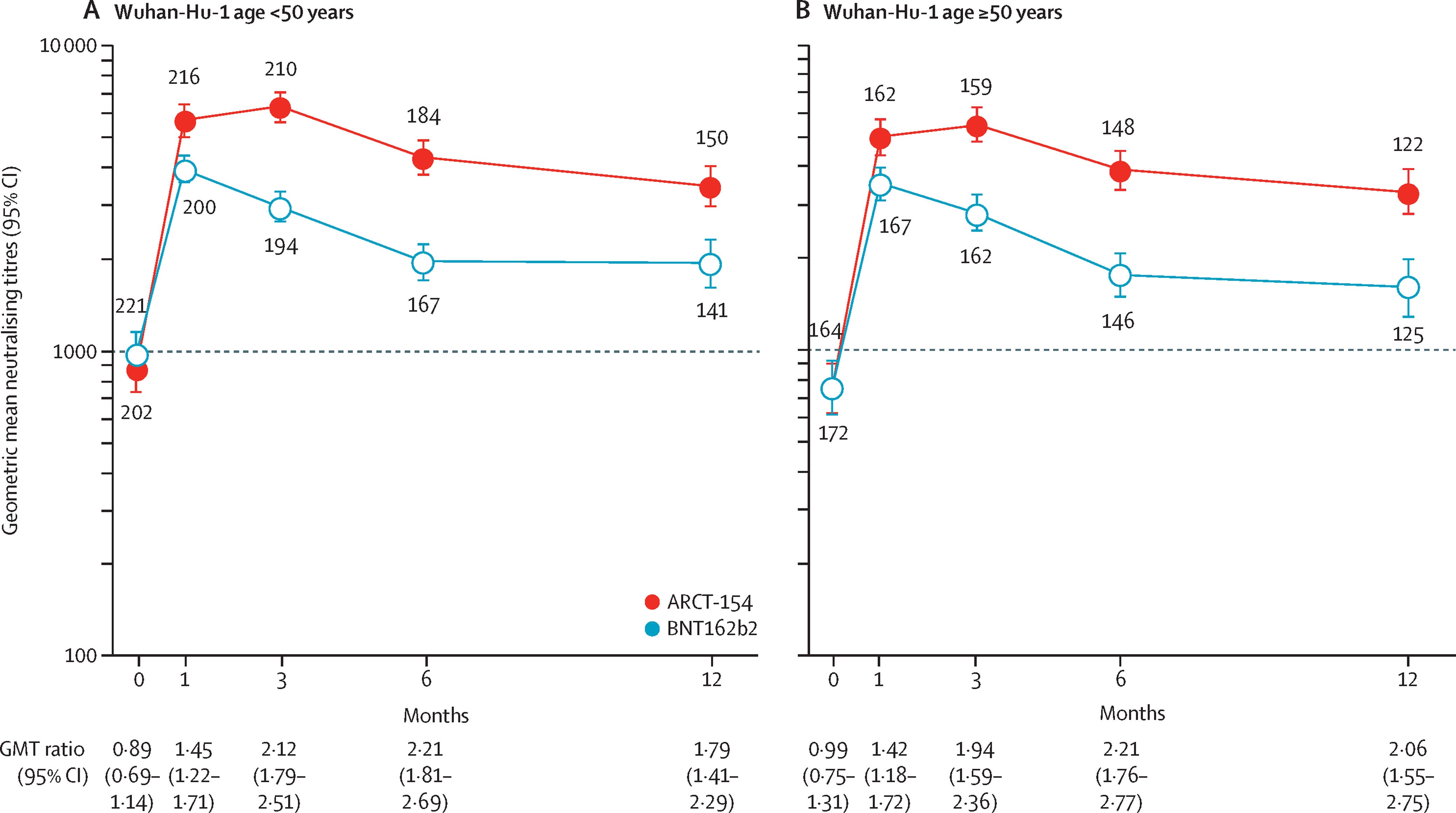
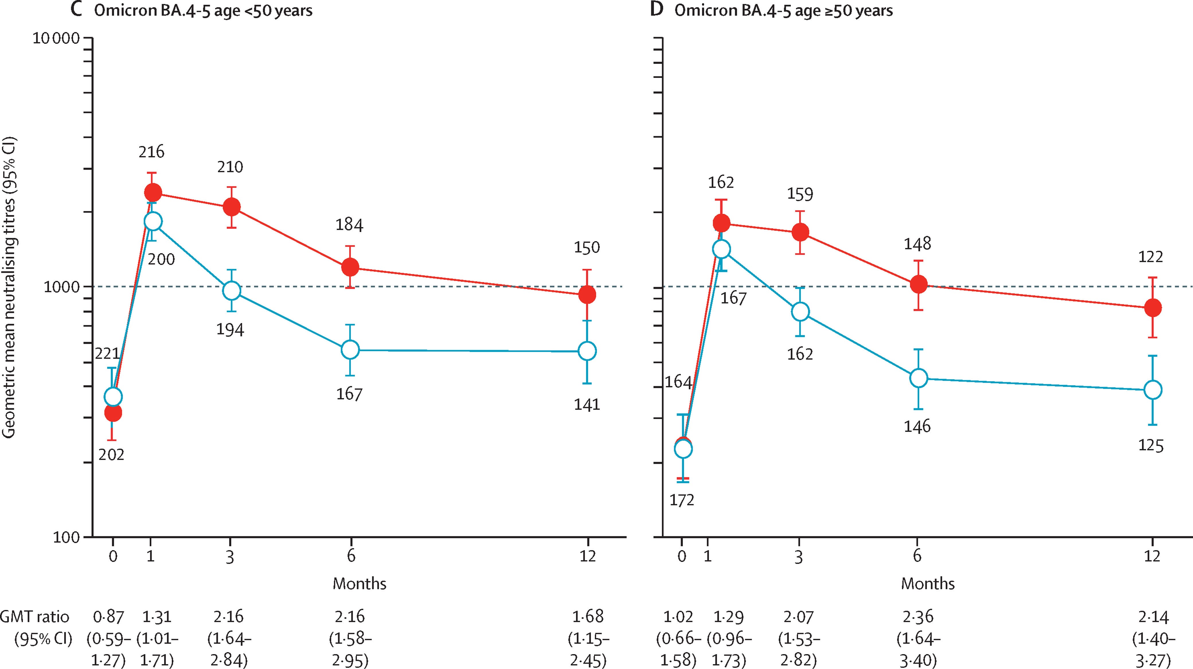
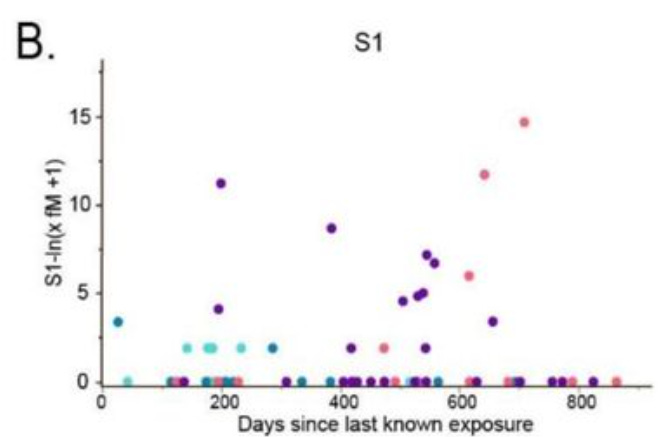


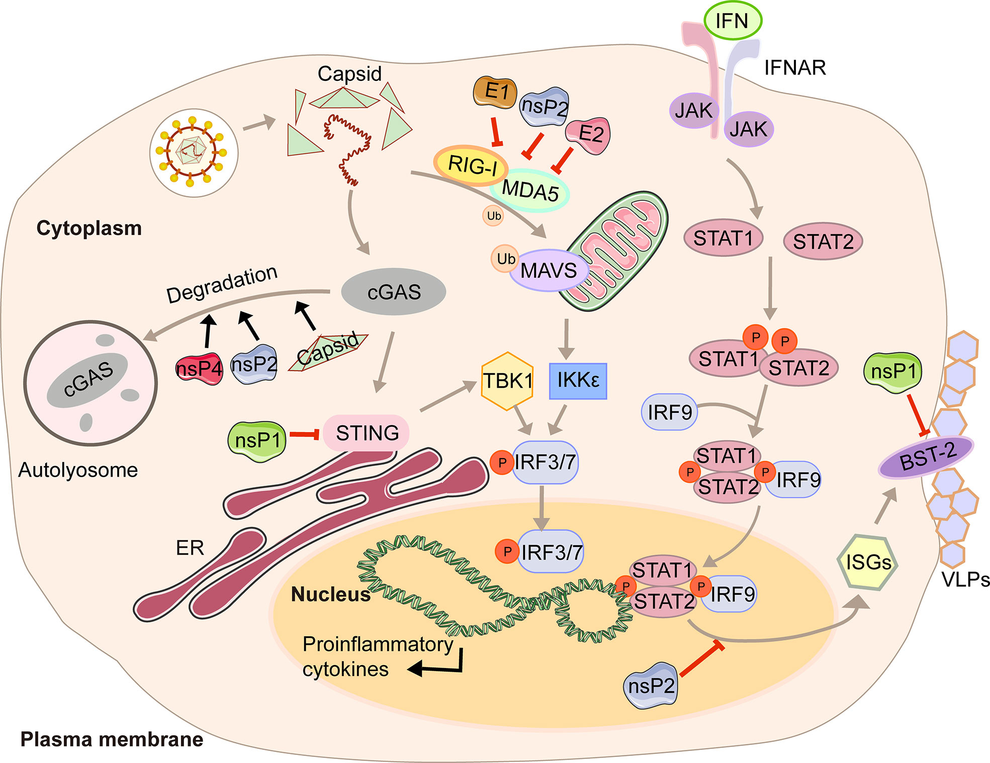


")


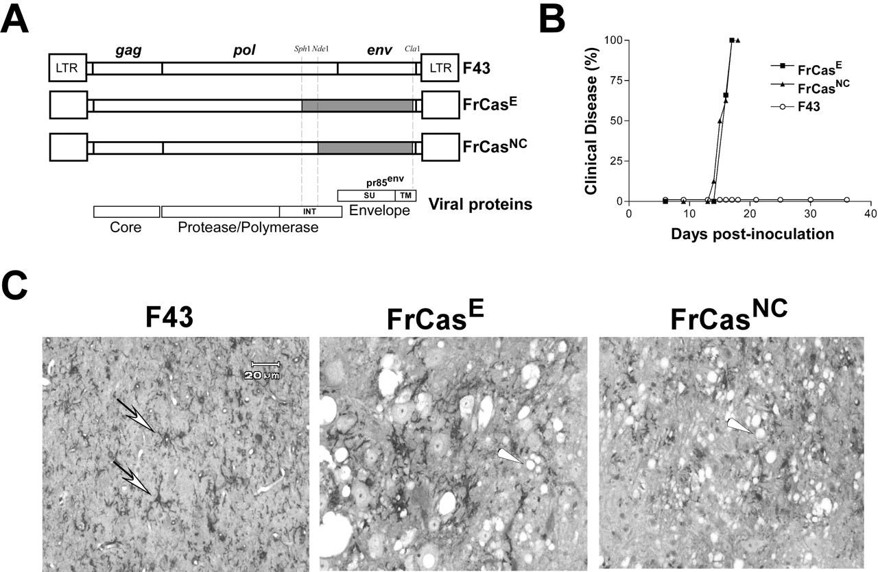


Wow, that was one incredible breakdown of how awful the self-replicating mRNA jabs are. Incredible work Doorless Carp! After this latest Yale study, which admits to finding the spike protein 709 days post jab, I was really hoping that the self-replicating versions would disappear, as they would be even more dangerous! I can only imagine how if we worried about shedding with the original jabs, how much worse this would be with self-replicating.
It should also be noted that USAID, is actually a CIA cut-out, so NewsGuard is obviously a wholly government agency controlled fact-checker and basically akin to that army regiment out of the UK that harasses people on X about everything covid.
Really really hoping that the MRNA jabs are withdrawn from the market and the RFKJr follows through on reducing all vaccines on the current schedule for children and adults. Very promising that he postponed, indefinitely, the thrice yearly vaccine meeting for boosting jabs to the US public! That includes the flu shots.
Cyprinus Carpio, another simply magnificent missive. We just digested Genervter's latest, and between the two of you, one can find the truth.
"The cure is worse than the disease. It’s a “feature” of replicon vaccines."
Indeed, not a bug.
"Blockbuster Yale Study: Millions Of Long COVID Patients Might Actually Be Vaccine Injured"
This was an expected outcome or feature which was an objective.
"Who needs to fully understand how something works before mass-vaccinating the world?"
Those with well defined goals and objectives.
"To the vaccinee, almost every alphavirus protein is working to suppress their immune system. It’s not the virus itself, but the immune response that causes the damage (plus adverse events)"
An objective, not happenstance.
Somebody just needs to tell the “experts” at the Mayo Clinic... Dr. Angela Rasmussen”
Many "expert" opinions are derived from a well funded payroll viz as opposed to the in vitro tests where lipid rafts are not present, the nature and functions of nuclear transport lipid rafts in vivo might yield different results. But they knew that in advance as well as...
"They aren’t interested in signs of cancer, teratogenicity, autoimmune disorders, vascular impairment, complex neurological disorders, immune suppression, class switching, or any long-term conditions..."
The above are ignored, because those are actual short and long term goals.
“Endoplasmic reticulum (ER) stress induced by a neurovirulent mouse retrovirus is associated with prolonged binding and retention of a viral protein in the ER... ER stress is linked to protein misfolding and accumulation."
If one wants amongst other things, a common element of viral infection viz lipid metabolome remodeling - to deleterious ends ( via PtdIns imbalance = ER stress), and all of the related consequences in a greatly accelerated rate across global populations...
One can achieve this by declaring pandemic, mandates for CV spike based mRNA vaccines, and cover all their tracks by blaming it on the virus.
"I wouldn’t trust them to put a plaster on my thumb"
These people aren't stupid, they have committed their acts with malice aforethought, and the ends justify the means Mr Mulder.
https://youtu.be/uki4lrLzRaU?&t=87